


Veterinary Guide to Eye Cancer in Dogs 2025 🐶
Dans cet article
Veterinary Guide to Eye Cancer in Dogs 2025 🐶
By Dr. Duncan Houston BVSc
🔍 Introduction
Eye tumors in dogs range from benign to aggressive malignant forms—such as melanomas, carcinomas, lymphomas, and mast cell tumors. Early detection and tailored treatment are vital to preserve vision and overall health. This 2025 guide provides veterinary insights into types, clinical signs, diagnostic workup, treatment plans, and prevention strategies. 🩺
💡 Common Types of Ocular Tumors
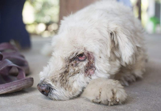
- Uveal melanoma: Most frequent intraocular tumor arising in iris, ciliary body, or choroid; may cause mass, pupil distortion, bleeding, uveitis, or glaucoma.
- Limbal (epibulbar) melanoma: Raised pigmented lesion at corneal-scleral junction, often with tearing or conjunctivitis.
- Meibomian gland adenoma/adenocarcinoma: Lumps on eyelid margin; adenomas are usually benign, adenocarcinomas rare and more aggressive.
- Mast cell tumors: Can appear on eyelids; need FNA/histopathology as they mimic skin lesions.
- Squamous cell carcinoma: Ulcerated masses, often in nonpigmented areas—photodamage suspected.
- Conjunctival or orbital tumors: Include lymphomas, adenocarcinomas, meningiomas, osteosarcoma; may cause swelling or bulging.
🚨 Clinical Signs to Watch For
- Visible mass—eyelid lump, dark spot on iris, or bulge in eyeball.
- Secondary effects: tearing, bleeding, cloudiness, squinting, eye bulging, pupil shape change.
- Vision impairment, possible uveitis or glaucoma—pain signs: blepharospasm, lethargy.
- Systemic signs are less common but may include weight loss or lethargy in aggressive tumors.
🔬 Diagnostic Strategy
- Eye exam with ophthalmoscope, slit-lamp, tonometry to evaluate the lesion and pressure.
- Ocular ultrasound, CT/MRI for depth, invasion, orbit assessment.
- FNA or biopsy to confirm histopathology and rule out mimics (cysts, abscesses).
- Thoracic/abdominal imaging to screen for metastasis in malignant cases.
- Bloodwork to assess general health and suitability for anesthesia.
🛠 Treatment Options
- Surgical excision: Eyelid tumors often removed with margins; rest assured long-term control vs cosmetic outcomes.
- Enucleation: Removal of eye for aggressive intraocular tumors, painful glaucoma, or when vision already lost.
- Radiation/laser therapy: Plaque brachytherapy, thermotherapy for uveal melanoma—reduces need for enucleation.
- Adjunctive therapy: Chemotherapy or immunotherapy for malignant or metastatic tumors.
- Supportive care: Topical antibiotics, anti-inflammatories, pain medication for underlying inflammation and comfort.
📈 Prognosis & Follow‑Up
- Benign eyelid tumors—excellent outcome after surgery.
- Limbal melanomas and uveal melanomas—usually low metastatic risk; prognosis depends on size and early treatment.
- Malignant types—guarded prognosis with risk of orbit invasion and metastasis.
- Regular monitoring: eye exams every 3–6 months post-treatment; imaging for systemic spread if malignant.
🛡 Prevention & Owner Advice
- Routine ocular exams—early detection improves outcomes.
- Protect from UV light—shade and doggy goggles like Rex Specs may help.
- Watch for new masses, swelling, or tearing—don’t delay vet exams.
- Breeding advice: avoid passing genetic cancer predispositions.
🔧 Recommended Tools & Services
- Ask A Vet App: 24/7 guidance on eye mass recognition, urgency, and referral 📱
✅ Final Thoughts
Eye cancer in dogs varies—some remain treatably benign, others demand aggressive intervention. With early evaluation, proper diagnostics, and tailored therapies, many dogs keep their vision and quality of life. Use Ask A Vet, expert support, into 2025 and beyond. 🐾❤️
Download the Ask A Vet app today to detect eye abnormalities, plan treatments, and support ocular healing. 📱💡


