

Vet Guide to Orbital Diseases in Dogs 2025 🐶🩺
In this article
Vet Guide to Orbital Diseases in Dogs 2025🐶🩺
By Dr. Duncan Houston BVSc
Orbital diseases affect the tissues around the eye—muscles, fat, nerves, vessels, and bone within the socket. They may be caused by infection, trauma, inflammation, or tumors, and often present as protruding (exophthalmos), sunken (enophthalmos), or misaligned eyes.
📍 Common Causes
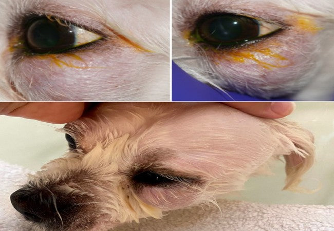
- Orbital cellulitis/abscess: Often from tooth root infections, grass awns or sinus disease. Signs include eyelid swelling, pain on mouth opening, protruding third eyelid, conjunctivitis, and exposure keratitis.
- Orbital neoplasia: Primary (adenomas, melanomas) or secondary tumors often cause gradually progressive exophthalmos, blindness, strabismus, and pain.
- Idiopathic orbital inflammation: Immune-mediated, may result in sudden swelling and discomfort; advanced imaging and biopsy are essential.
- Muscle-related inflammation: Orbital myositis or masticatory muscle myositis (MMM) can also push the eye forward, causing pain on chewing and third eyelid protrusion.
- Trauma & hemorrhage: Blunt injuries and bleeding within the orbit lead to sudden exophthalmos; globe replacement or enucleation may be needed.
⚠️ Clinical Signs
- Protruding (exophthalmos) or sunken (enophthalmos) eye(s), often with third eyelid prolapse, strabismus, anisocoria or impaired eye movement.
- Pain on opening the mouth or palpating the orbit, corneal ulcers (due to exposure), tearing or discharge.
- Systemic signs: lethargy, fever (infection), neurological deficits or vision loss in tumor cases.
🔬 Diagnostics
- Physical exam: Globe retropulsion shows limited movement with space-occupying lesions; pain on mouth movement suggests cellulitis or MMM.
- Imaging (CT/MRI): Key for evaluating extent, differentiating inflammation vs tumor and guiding biopsies.
- Ultrasound & radiography: Less detailed, but useful for initial assessment of fluid, foreign bodies, bone or tooth issues.
- Biopsy/FNA & histopathology: Essential for diagnosing neoplasia and idiopathic inflammation.
- Blood tests: Assess for infection or immune-mediated disease when evaluating systemic involvement.
💊 Treatment Options
- Cellulitis/abscess: Drainage via oral approach, systemic antibiotics, warm compresses, topical eye protection; prognosis is good with prompt therapy.
- Idiopathic inflammation: Immunosuppressive doses of corticosteroids ± adjuncts; long-term monitoring due to relapse.
- Tumors: Surgical excision with or without enucleation, radiation or chemotherapy for malignant cases; prognosis varies and is often guarded.
- Orbital myositis/MMM: Long-term corticosteroids; MMM may require physiotherapy and soft diets for jaw mobility.
- Trauma: Globe replacement and tarsorrhaphy for proptosis; enucleation if globe is non-viable.
📈 Prognosis & Monitoring
- Infections and abscesses: usually resolve fully within weeks with treatment.
- Idiopathic inflammation: variable - may respond quickly to steroids, but relapses are common.
- Tumors: early removal helps, but malignancies often have guarded to poor outcomes.
- Trauma/proptosis: globe salvage ~40% in some cases; lifelong ocular care often needed.
- Monitor via exams and repeat imaging to check for recurrence, complications (ulcers, dry eye) or treatment reactions.
✅ Dr Houston’s Clinical Tips
- 🔍 Always assess retropulsion & jaw pain—cellulitis often mimics dental issues.
- 📸 Use advanced imaging early—orbital signs are often hidden behind eyelids.
- ⚖️ Choose treatment tailored to the cause: antibiotics for infection, steroids for inflammation, surgery for tumors.
- 🛡️ Protect corneas with lubricants or tarsorrhaphy if eyelid closure is impaired.
- 📆 Schedule follow-up visits even after resolution—relapse and secondary ulcers are common.
If your dog has a bulging or sunken eye, pain around the jaw or mouth, swelling, or vision loss, seek veterinary care immediately. With prompt diagnosis and targeted therapy, many dogs can recover comfort, vision, and quality of life. 🐾❤️


