

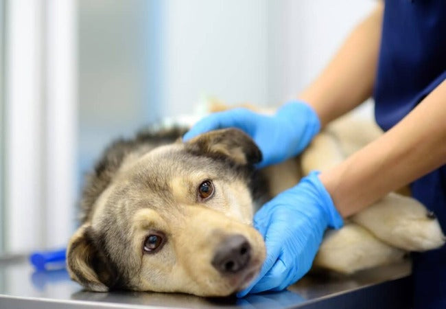
Esophageal Strictures in Dogs – Vet‑Led Guide 2025 🐶🍽️
In this article
Esophageal Strictures in Dogs – Vet‑Led Guide 2025 🐶🍽️
By Dr. Duncan Houston BVSc
Hello, I’m Dr Duncan Houston, BVSc, founder of Ask A Vet. When a dog develops an esophageal stricture—a narrowing of the swallowing tube—it becomes a challenge for them to eat and drink. In this in-depth 2025 guide, we’ll cover what causes strictures, how to recognize the signs, the best diagnostic tools, treatment options like balloon dilation and B‑tubes, feeding strategies, and long-term outlooks. Let’s restore that happy, worry-free mealtime together. 🍽️🐕
📘 What Is an Esophageal Stricture?
An esophageal stricture is a narrowing of the esophagus caused by scar tissue formation after injury. Unlike megaesophagus, where the esophagus itself dilates, strictures create a blockage that impedes normal swallowing. Common culprits include damage from anesthesia, foreign bodies, acid reflux, caustic medications, or inflammatory injury.
🚩 Causes & Risk Factors
- Foreign body lodgment: bones, toys stuck in the esophagus can erode the lining.
- Anesthetic reflux: gastric acids can damage the esophageal lining during anesthesia.
- MEDICATION-INDUCED: Doxycycline or clindamycin tablets are causing caustic mucosal injury.
- Chronic vomiting/reflux: acid-peptic injury leads to fibrosis over time.
- Caustic substances: ingestion of chemicals or radiation exposure.
- Neoplasia: uncommon—tumors or parasites like Spirocerca lupi may narrow the lumen.
🚨 Who Is Most Affected?
Dogs of any age or breed can develop strictures. However, risk increases after incidents with anesthesia, ingesting foreign bodies, or receiving caustic pills. Repeated episodes of vomiting or reflux push the esophagus toward scarring.
🩺 Clinical Signs & Red Flags
- Reluctance or difficulty swallowing food or water.
- Immediate regurgitation after eating—often undigested or dry food.
- Painful swallowing, drooling, gagging, or coughing.
- Loss of appetite, weight loss, and dehydration.
- Soft or liquid food may pass better than kibble.
- Possible aspiration pneumonia—look for coughing or respiratory distress.
🔍 Diagnostic Approach
- History & physical exam: note recent anesthesia, foreign body removal, vomiting events, and physical signs.
- Bloodwork and radiographs: evaluate inflammation, rule out pneumonia or tumors.
- Contrast esophagram or fluoroscopy: barium swallow helps identify location, length, and severity, but be cautious of aspiration.
- Endoscopy: direct visualization; can optionally proceed with balloon dilation during the same procedure.
- Biopsy: mucosal sampling helps rule out cancer in atypical or non-responsive cases.
💉 Treatment Modalities
1. Balloon Dilation
- Preferred first-line treatment; 70–88% success rates.
- Performed 2–4× at 5–10-day intervals under anesthesia.
- Tissue pressure breaks scar tissue gradually; larger balloons are used each session.
- Risks include esophageal tearing (4–9%), perforation, bleeding, and pneumonia.
2. Bougienage / Guidewire Dilation
An alternative, manually-guided dilation is used less frequently due to injury risk, but effective in many clinics.
3. B‑Tube (Balloon Esophagostomy Feeding Tube)
- Tube placed after initial dilation; balloon inflated daily at home to maintain lumen.
- Dim risks of repeated anesthesia; dogs often tolerate gag reflex without tears.
- Complications include regurgitation, site infection, and occasional recurrence.
4. Surgical Resection
Rarely indicated—only for long strictures, tumors, or those not responding to dilation. Involves segmental resection and re-anastomosis under general anesthesia.
5. Medical Management
- Proton pump inhibitors or antacids post-dilation or for mild strictures.
- Bland or liquid diets to reduce further mucosal injury.
- Nutritional support—temporary feeding tubes if swallowing remains inadequate.
🏡 Home Care & Feeding Strategies
- Warm, soft or pureed food is best tolerated.
- Feed small portions slowly—allow time between mouthfuls.
- Elevate bowls to minimize aspiration risk.
- Posture dogs upright after meals for 5–10 minutes.
- Monitor for coughing, choking, weight changes—log issues.
- Administer antacids or PPIs as recommended.
- If a B‑tube is in place, follow strict inflation protocols.
- Re-check the vet regularly for scopes or imaging to confirm healing.
📅 Prognosis & Long-Term Outlook
- Early-detected stricture treated promptly has a good to excellent prognosis.
- 70–85% regain functional swallowing, though with modified diets.
- Severe, long strictures have guarded prognoses—even with multiple dilations.
- Recurrence is common—repeat procedures may be necessary; B‑tube improves maintenance.
- Complications like perforation and pneumonia require careful monitoring.
- With routine care and monitoring, many dogs lead comfortable lives post-treatment.
🐾 Role of Ask A Vet
Need immediate feeding guidance or monitoring questions? Connect 24/7 via Ask A Vet. Post-dilation feeding aids like raised bowls, pureed food mixers, and B‑tube supplies are available. 💙
✨ Key Takeaways
- Esophageal strictures are caused by scar tissue narrowing the esophagus after damage.
- Symptoms include drooling, gagging, regurgitation, weight loss, and coughing.
- Diagnosis relies on contrast imaging, endoscopy, and biopsies when needed.
- Balloon dilation is the gold standard; B‑tubes help sustain healing.
- Home feeding adjustments and medications support recovery.
- Most dogs restore functional swallowing, though many need dietary changes.
- Early intervention and consistent follow-up provide the best outcomes—don’t hesitate to reach out to your vet or Ask A Vet. 🍽️❤️
If your dog struggles to swallow or frequently regurgitates—especially after anesthesia or ingesting something—they may have an esophageal stricture. Contact your veterinarian or Ask A Vet promptly. Early treatment gives your dog the best chance to enjoy mealtime again. 🩺


