

Non‑Ulcerative Keratitis in Dogs – Vet Guide 2025 🐶👁️🩺
In this article
Non‑Ulcerative Keratitis in Dogs – Vet Guide 2025 🐶👁️🩺
By Dr. Duncan Houston BVSc
Hello, I’m Dr Duncan Houston, BVSc, founder of Ask A Vet. When your dog’s corneal surface becomes inflamed without ulceration, it can cause discomfort and vision issues if untreated. This comprehensive 2025 guide covers types, causes, diagnostics, medical and surgical treatments, home care tips, and long‑term strategies to preserve eye health and comfort. Let’s help your dog see clearly and live fully! 🐾
📘 What Is Non‑Ulcerative Keratitis?
Non-ulcerative keratitis is inflammation of the cornea where the epithelial surface remains intact—there's no ulceration but there may be cloudiness, pigmentation, vascularization, or discharge. Unlike ulcerative forms, no fluorescein dye uptake occurs.
🧩 Types & Breed Predispositions
- Infectious keratitis: non-ulcerative bacterial, viral, or fungal infections.
- Chronic Superficial Keratitis (Pannus): immune-mediated, often in German Shepherds, Huskies, Greyhounds—characterized by pigmentation & vessels.
- Pigmentary keratitis: common in brachycephalic breeds (Pugs, Shih Tzus), due to chronic exposure or irritation.
- Keratoconjunctivitis sicca (KCS): dry-eye causing chronic surface inflammation, common in Cocker Spaniels.
- Exposure/trauma-related: mechanical damage, chemical injury, or trichiasis.
🚩 Clinical Signs
- Cloudy, hazy, or brown‑colored cornea.
- Growth of blood vessels (vascularization) into cornea.
- Discharge—clear, mucopurulent or yellowish.
- Squinting, redness (blepharospasm, conjunctival hyperemia).
- Subtle systemic signs—lethargy, reduced appetite, vision impairment.
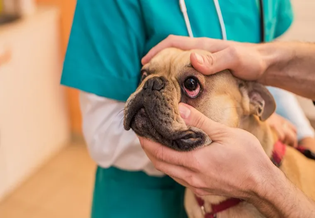
🔍 Diagnostic Approach
- History & exam: breed at‐risk, recent trauma, duration of signs.
- Fluorescein stain: essential to confirm non‐ulcerative nature—no stain uptake present.
- Schirmer tear test: assess tear production—essential for KCS cases.
- Corneal culture/cytology: sample any discharge to identify infection, especially for bacterial/fungal causes.
- Intraocular pressure check: screen for glaucoma or anterior uveitis.
- Conformational check: evaluate eyelid/re‑lacion anatomy, eyelashes, globe size.
- Immunologic/biopsy: in chronic pannus to confirm immune etiology; fluorescein angiography or biopsy may be required.
💉 Medical Treatments
- Topical antibiotics: e.g., ofloxacin, moxifloxacin—if infection suspected.
- Topical corticosteroids: prednisolone acetate or dexamethasone for inflammation—avoided if ulceration is present.
- Topical cyclosporine or tacrolimus: for KCS and pannus—modulates immune response.
- Lubricants/artificial tears: used frequently in KCS, pigmentary keratitis.
- Oral NSAIDs or systemic steroids: short course in severe inflammation.
⚠️ Surgical & Advanced Interventions
- IPL or CO₂ laser: in recalcitrant pannus to reduce vessels/pigmentation.
- Excision of trichiasis/entropion: eyelid surgery corrects mechanical damage.
- Corneal cryotherapy/grafting: in chronic non‐healing pannus/scar tissue.
- Nictitating membrane flap: protective bandage for deep ulcerative cases—less relevant here, but may aid severe inflammatory corneal conditions.
🏡 Home Care & Long-term Management
- Medication adherence: vital for chronic cases like pannus and KCS—non-compliance leads to recurrence.
- UV/air irritant protection: dog goggles or indoor shelter—especially in pannus and pigmentary keratitis.
- Regular cleaning & lubrication: warm wet compresses before drops help enhance penetration.
- Monitor vision & discomfort: note squinting, blinking, or pawing at eyes; adjust environment accordingly.
- Periodic recheck: every 3–6 months to tailor therapy, reduce flare-ups, reference veterinary ophthalmologist if unresponsive.
📅 Prognosis & Follow‑Up
- KCS & pannus: lifelong control—excellent prognosis with maintenance therapy.
- Pigmentary keratitis: often slowly progressive—lubrication and lens protection help; may plateau with chronicity.
- Infection-related: good to excellent with appropriate antibiotics; culture helps guide therapy.
- Mechanical or trauma: complete resolution after surgical correction in most cases.
- Vision preservation: early detection and treatment are key—untreated cases may result in blindness.
🐾 Support with Ask A Vet
- Need guidance on eye-drop schedules or swelling? Connect instantly with a vet through Ask A Vet.
✨ Key Takeaways
- Non-ulcerative keratitis is corneal inflammation without ulceration—presenting as cloudiness, pigment, vessels or discharge.
- Common causes include immune-mediated pannus, pigmentary keratitis in flat-faced breeds, dry eye, infections, and mechanical irritation.
- Diagnose via fluorescein staining, Schirmer tear test, culture, intraocular pressure, and possibly biopsy.
- Treatment varies—antibiotics, steroids, immunosuppressives, lubrication, and surgical corrections.
- Lifelong home care (drops, UV protection, cleaning) greatly improves outcomes.
- With early intervention and ongoing support, dogs can maintain comfort and clear vision. 🐶❤️
If you notice cloudiness, discoloration, squinting or discharge in your dog’s eyes, don’t delay—contact your vet or reach out to Ask A Vet. Timely treatment protects vision and comfort. 🩺


