


Pythiosis in Dogs 2025: Vet Guide to Diagnosis, Treatment & Care 🩺🐾
In this article
Pythiosis in Dogs 2025: Vet Guide to Diagnosis, Treatment & Care 🩺🐾
By Dr. Duncan Houston BVSc
Hi, I’m Dr Duncan Houston BVSc, veterinarian and founder of Ask A Vet. In this detailed 2025 guide, we explore pythiosis—a serious, life-threatening disease caused by the water mold Pythium insidiosum. It primarily affects dogs exposed to stagnant water and soil, presenting as gastrointestinal (GI) masses or draining skin ulcers. We'll cover signs, testing, treatment options, prognosis, and prevention. Discover how Ask A Vet, tools support you and your pup through this journey. 💙🐶
1. What Is Pythiosis?
Pythiosis is caused by the oomycete Pythium insidiosum, a water mold—not a true fungus—that infects dogs through wounds or ingestion of contaminated water. It causes aggressive granulomatous disease, primarily affecting the GI tract or skin/subcutaneous tissues. Systemic spread can occur .
2. Where & Why Dogs Get It ⚠️
- Primarily in tropical/subtropical regions—Gulf Coast, Southeastern US, parts of California, Arizona, Wisconsin, and globally in Brazil, Southeast Asia, and Australia .
- Exposure stems from swimming or drinking in stagnant water (ponds, swamps, bayous) .
- Young, large-breed, active dogs—German Shepherds, Labradors, Cavaliers—are at higher risk .
3. Forms & Clinical Signs 🕵️♂️
3.1 Gastrointestinal (GI) Pythiosis
- Chronic vomiting, diarrhea (often bloody), weight loss, reduced appetite, and fever .
- Firm abdominal masses—especially near stomach, duodenum, ileocolic junction; palpable on the exam .
- Ultrasound/CT shows thickened intestinal walls, loss of layering, lymphadenopathy .
- Slow progression; dogs often decline only after months despite relatively normal early behavior .
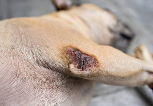
3.2 Cutaneous/Subcutaneous Pythiosis
- Non-healing skin nodules or ulcers (legs, tail, abdomen, perineum) with draining sinus tracts .
- Serosanguinous or purulent discharge, necrosis, and granulation tissue may be present .
- Often mistaken for other infections or neoplasia, leading to diagnostic delays .
4. How Is It Diagnosed? 🔬
- Clinical suspicion: history of water exposure + GI mass or non-healing ulcer raises suspicion .
- Imaging: ultrasound or CT to identify lesions and guide biopsies .
- Histopathology: biopsies with granulomatous inflammation; hyphae best seen with Gomori methenamine silver or PAS stain .
- Serology/ELISA: anti-Pythium IgG assay is highly sensitive and specific (>99%) and useful for monitoring .
- Culture/PCR: tissue culture and molecular testing to confirm species .
- Routine labs: nonspecific findings—anemia, eosinophilia, hypoalbuminemia .
5. Treatment Strategies 🛠️
5.1 Surgical Removal
- Wide surgical excision is essential for cutaneous lesions—may require amputation .
- GI lesions often require segmental bowel resection with clean margins .
- Complete removal offers the best chance for remission, but recurrence is possible .
5.2 Medical Therapy
- Combination antifungals (itraconazole + terbinafine ± amphotericin B) administered for ≥6–12 months .
- Given Pythium lacks ergosterol, medical therapy alone rarely cures—but may support surgical outcomes or control GI disease .
- Prednisone is used to reduce inflammation and improve antifungal penetration .
5.3 Immunotherapy
- Antigen-based immunotherapy (MiraVista, PAVLAB) stimulates immune response; shows promise especially combined with surgery .
6. Monitoring & Prognosis 📊
- GI form: guarded prognosis; survival improves with surgery + meds, ~75% remission .
- Cutaneous form: prognosis is better when full excision is achieved; untreated cases often fatal .
- Regular monitoring with serial serology and imaging helps track response .
- Recurrence risk exists—long-term surveillance is essential.
7. Prevention Tips 🏡
- Avoid letting dogs swim in stagnant water areas; provide clean drinking water .
- Clean and dry skin wounds promptly after outdoor exposure .
- Regular veterinary check-ups for at-risk dogs during high-risk seasons.
- Maintain strong nutrition and overall health—immune function plays a key role .
8. Support from Ask A Vet 💡
- Ask A Vet: Telehealth triage, pre-surgery guidance, lab interpretation, and follow-ups.
9. When to Contact the Vet Immediately 🚨
- Persistent vomiting, diarrhea, palpable abdominal mass.
- Non-healing draining skin wound or ulcer.
- Weight loss despite eating and meds.
- Confirm diagnosis or initiate antifungal/immunotherapy.
10. Final Thoughts 📝
Pythiosis is a serious water‑mold infection that requires fast, aggressive treatment combining surgery, long-term meds, and often immunotherapy. While prognosis remains guarded, early recognition and comprehensive care significantly improve outcomes. Tools from Ask A Vet, help you navigate treatment, monitor recovery, and stay proactive in 2025 and beyond. 🐾💙
If your dog has suspicious symptoms or skin lesions after water exposure, book a telehealth consult via AskAVet.com. Download our app to track treatments, symptoms, and follow-up care. 🌟


