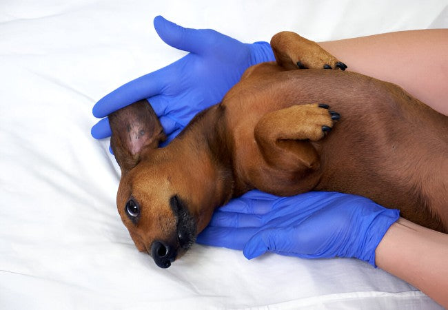
Testicular Tumors in Dogs 2025: Vet Guide to Detection & Care 🐾

In this article
Testicular Tumors in Dogs 2025: Vet Guide to Detection & Care 🐾
By Dr. Duncan Houston BVSc
Testicular tumors are among the most frequent reproductive tumors in intact male dogs, particularly affecting older dogs and those with undescended (cryptorchid) testicles. This guide explains tumor types, signs, diagnostics, treatments, and supportive care informed by current veterinary practices.
1. 📊 How Common Are They?
Testicular tumors occur in about 4–7% of male dog tumors, with up to 27% of intact males developing them in their lifetime. Cryptorchid dogs are at much higher risk—studies show a 13‑fold increase.
2. 🧬 Types of Testicular Tumors
- Interstitial (Leydig) cell tumors: Most common (~51%), typically benign and asymptomatic.
- Seminomas: ~24%; usually benign, can occur in cryptorchids, occasionally estrogen-secreting.
- Sertoli cell tumors: ~18%; may produce estrogen causing hyperestrogenism—feminization, marrow suppression; higher metastatic tendency.
- Mixed tumors: ~7%; combination of types.
3. ⚠️ Who’s at Risk?
- 📅 Age: Typically 7+ years; risk increases over 10 years.
- 🐕 Breed predisposition: Boxers, German Shepherds, Afghan Hounds, Weimaraners, Shetland Sheepdogs, Collies, Maltese, Labs, Goldens, Yorkies.
- 🔝 Cryptorchidism: retained testicles drastically increase risk.
4. 🔍 Typical Signs to Watch
- Scrotal enlargement or asymmetry.
- Unilateral or bilateral testicular masses on palpation.
- Feminization in Sertoli tumors: mammary enlargement, nipple prominence, hair loss, hyperpigmentation, and squatting to urinate.
- Loss of fertility in breeding males.
- Advanced cases: weight loss, lethargy, loss of appetite, or signs of metastasis.
5. 🩺 Diagnostic Approach
Early detection often begins during routine exams. Diagnostics may include:
- Physical examination and scrotal palpation.
- Ultrasound to locate and characterize lesions, especially in cryptorchids.
- Fine-needle aspirate or biopsy gives preliminary cytology.
- Post-neuter histopathology confirms tumor type and informs prognosis.
- Blood work and urinalysis, especially if estrogen effects are suspected.
- Chest X‑rays/abdominal imaging for metastasis in malignant cases.
6. 🛡️ Treatment Options
• Neutering (Orchiectomy)
The gold-standard treatment is surgical removal of affected testicles (and retained ones). This often cures the disease and eliminates estrogen-producing tumors.
• Adjuvant Therapy for Metastasis
Cancer spread to lymph nodes or beyond may require chemotherapy protocols (e.g., platinum-based) or radiation, a rare but documented use.
• Surveillance Only
Small, benign-appearing tumors in older dogs may be monitored until they grow or cause problems.
7. 🏥 Prognosis & Outcome
- Overall, excellent when localized and treated early.
- Sertoli and seminoma spread risk is <15%, and interstitial virtually none.
- Metastatic disease has a guarded prognosis—early detection improves odds.
8. 🏡 Post-Surgery Support
- Monitor the incision daily—look for swelling, discharge, redness.
- Use an Elizabethan collar to prevent licking, especially in sensitive dogs.
- Pain management per vet instructions.
- Light leash walks until sutures dissolve or are removed (~10–14 days).
- Gradually resume normal activity over 4–6 weeks.
- Access Ask A Vet App anytime for concern or guidance.
9. 🌱 Long-Term Monitoring & Prevention
- Neuter early to prevent the development of testicular tumors.
- Perform routine scrotal palpation on intact males, especially older or cryptorchid.
- Consider genetic counseling and avoid breeding cryptorchids.
- Regular wellness exams with blood tests for hormone or organ function.
10. 🔍 Cryptorchidism & Its Risks
Cryptorchid dogs carry a significantly higher risk, particularly for Sertoli and seminoma tumors.
Standard care is surgical removal of both retained and normal testes to prevent future tumors and eliminate heat cycle behavior.
11. 📌 Summary for Dog Parents
- Testicular tumors are common in intact males, especially older and cryptorchid dogs.
- Watch for scrotal enlargement, lumps, feminization signs, and systemic illness.
- Diagnosis includes physical exam, imaging, cytology, and histopathology.
- Neutering cures most, with adjunct therapies reserved for spread.
- Early detection and surgery offer an excellent prognosis.
- Post-op care and monitoring with Ask A Vet,


