

Vet Guide to Corneal Degenerations & Infiltrations in Dogs 2025 🐶🩺
In this article
Vet Guide to Corneal Degenerations & Infiltrations in Dogs 2025 🐶🩺
By Dr. Duncan Houston BVSc
Corneal degeneration involves deposits of lipid or calcium in the corneal epithelium or stroma secondary to local or systemic disease, while corneal dystrophy is an inherited primary disorder. Both may impact vision and comfort and require veterinary evaluation.
📍 Causes & Types
- Lipid degeneration: Fatty deposits due to lipid metabolism disorders (e.g., hypothyroidism, Cushing’s, diabetes, dyslipidemia); often exacerbated by prior corneal inflammation or ulcers.
- Calcific degeneration: Calcium deposition in older dogs or after chronic corneal disease/trauma; may ulcerate or cause discomfort.
- Corneal dystrophy: Genetic opacity of epithelial, stromal (lipid), or endothelial layers; non-inflammatory but may lead to edema, bullae, and ulcers.
- Pannus (Chronic Superficial Keratitis): Immune-mediated vascular and pigment infiltration, often photo-exacerbated; classic in German Shepherds and related breeds.
⚠️ Clinical Signs
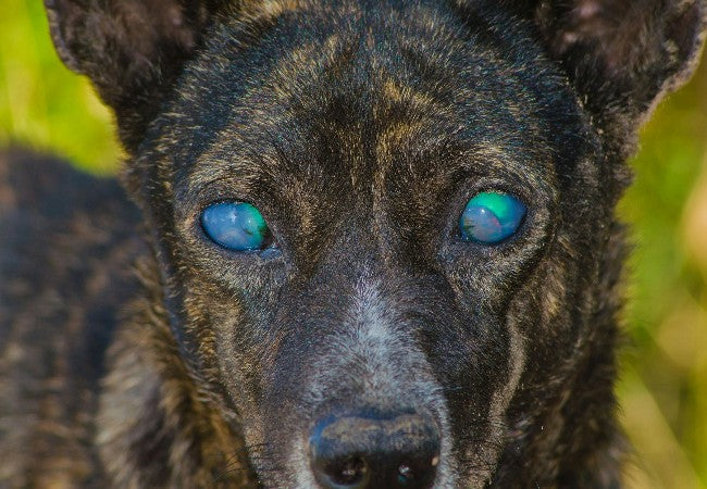
- White, yellow, or crystalline corneal spots; cloudiness or blue haze in endothelial cases.
- Possible corneal vascularization, redness, discharge, ocular discomfort if the degeneration ulcerates.
- Pannus exhibits early limbal blood vessels, pigmentation, and surface immune proliferation.
- Endothelial dystrophy leads to fluid-filled bullae and potential pain or ulceration.
🔬 Diagnosis & Testing
- Slit‑lamp examination to define lesion layer and opacity type.
- Fluorescein stain to detect any ulceration, especially in calcific and endothelial cases.
- Schirmer tear testing and tonometry for concurrent dry eye or pressure issues in immune disease.
- Systemic work-up (cholesterol, calcium, endocrine profiles) for metabolic infiltrations.
- Breed and age clues, plus family history, support inherited dystrophy diagnosis.
💊 Treatment & Care
- Lipid degeneration: Address systemic disease (diet, endocrine therapy); lubricant drops and monitor for ulcers.
- Calcific degeneration: Apply EDTA topical drops to chelate calcium; manage ulcers with antibiotics and pain control.
- Dystrophy: Superficial dystrophies typically only need monitoring; endothelial involvement is managed with hypertonic saline, topical anti-inflammatories, contact lenses, or keratoplasty for severe cases.
- Pannus: Lifelong topical immunomodulators (cyclosporine, tacrolimus) or steroids, plus UV protection with doggles or tinted training areas.
📈 Prognosis & Monitoring
- Lipid/calcific degenerations may stabilize or improve with systemic therapy; potential ulcer risk remains.
- Dystrophy is often benign with slow progression; endothelial cases require ongoing management.
- Pannus is controlled effectively with treatment, but flare-ups are common—UV avoidance is essential.
- Regular rechecks (every 6–12 months) to monitor lesion progression, comfort, and vision.
✅ Dr Duncan Houston’s Vet Tips
- 🔍 Investigate any corneal cloudiness—early differentiation avoids complications.
- 📊 Run lipid and calcium panels in senior dogs with corneal infiltrates.
- 🎯 Use EDTA tokens for calcific degeneration to chelate calcium.
- 🛡 Protect pannus-prone breeds with doggles and topical immunosuppression.
- 🚫 Avoid breeding dogs with dystrophy or pannus to limit inherited disease.
If your dog’s corneas show spots, haze, vessels, or discomfort—especially in predisposed breeds—book an eye check via AskAVet.com.🐾❤️


