

Vet Guide to Corneal Dystrophies in Dogs 2025 🐶👁️
In this article
Vet Guide to Corneal Dystrophies in Dogs 2025 🐶👁️
By Dr. Duncan Houston BVSc
Corneal dystrophies are inherited, bilateral, non‑inflammatory conditions that cause corneal cloudiness due to epithelial, stromal, or endothelial layer dysfunction. These deposits or fluid changes rarely cause pain but may affect vision in advanced stages. Diagnosing the type guides management and referral decisions. 💡
📍 Types of Corneal Dystrophy
- Epithelial: White/gray plaques or rings on surface; rarely erosive. Vision preserved.
- Stromal (lipid): Crystalline or doughnut-shaped fat or cholesterol deposits—typically asymptomatic.
- Endothelial: Fluid leaks causing bluish edema, bullae (blisters), and ulceration—most concerning for comfort and vision.
⚠️ Breed Predisposition & Onset
Common breeds include Beagles, Cocker Spaniels, Cavaliers, Huskies, Boston Terriers, Chihuahuas, Dachshunds, Shelties, and more.
👀 Signs & Impacts
- Variable corneal cloudiness—white, gray, silver or blue haze.
- Usually non‑painful, though endothelial forms may blister and ulcerate.
- Vision often unaffected unless changes are diffuse or ulcerative.
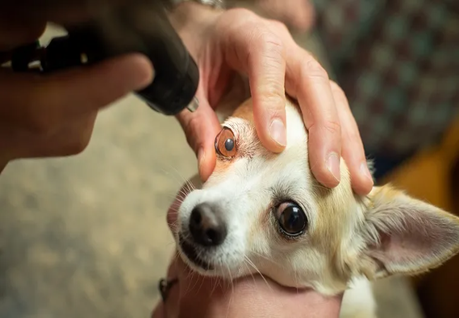
🔬 Diagnostic Approach
- Slit‑lamp or ophthalmoscopic exam to locate the affected layer.
- Fluorescein stain to detect ulcers (especially in epithelial/endothelial cases).
- Tonometry to rule out glaucoma; tear test for ocular surface disorders.
- Bloodwork if systemic lipid or calcium disorders suspected.
💊 Management & Treatment
- Epithelial/stromal forms usually need no treatment beyond monitoring.
- Endothelial cases may benefit from hypertonic saline, topical anti‑inflammatories, and bandage contact lenses.
- Refractory ulcers may require superficial keratectomy, conjunctival grafts, or transplant; repeat episodes are common.
- Treat systemic hyperlipidemia or hypercalcemia if present in stromal degeneration.
- Emerging therapies: ROCK inhibitors like netarsudil show promise for endothelial dystrophy, though still limited in availability.
📈 Prognosis & Follow‑up
- Superficial lesion prognosis is excellent; vision remains intact.
- Endothelial dystrophy management prevents ulceration; vision may decline if untreated.
- Annual ophthalmic exams are recommended for early detection and intervention.
✅ Dr Houston’s Clinical Tips
- 🔍 Monitor cloudiness—note changes or new discomfort.
- 😊 Reassure owners—most dystrophies are harmless and stable.
- 🚨 Act fast on signs of ulcers—bulging, redness, squinting—refer to an ophthalmologist.
- 🧬 Advise against breeding affected dogs—even mild dystrophy signals heritable disease.
If your dog’s eyes have cloudiness, crystals or blisters—especially in predisposed breeds—seek an eye exam via AskAVet.com.Early detection preserves both comfort and clarity of vision. 🐾❤️


