

Vet Guide to Corneal Dystrophies in Dogs 2025 🐶👁️
In this article
Vet Guide to Corneal Dystrophies in Dogs 2025 🐶👁️
By Dr. Duncan Houston BVSc
Corneal dystrophies are inherited, typically bilateral, non-inflammatory eye conditions where the cornea becomes cloudy due to material deposits or cellular dysfunction. They affect vision depending on the layer and severity. This guide explains the three major types—epithelial, stromal, and endothelial—along with breed predisposition, clinical signs, diagnosis, management, prognosis, and prevention based on veterinary best practice.
📍 Types of Corneal Dystrophy
- Epithelial dystrophy: Superficial white or gray rings/spots; sometimes painful if erosions develop. Vision is usually normal.
- Stromal dystrophy: Fat or lipid deposits cause gray, crystalline or doughnut-shaped spots in the stroma—vision is typically unaffected unless extensive.
- Endothelial dystrophy: Inner cornea fluid retention leads to cloudiness, bullae, and ulcers—can be painful and may impair vision.
⚠️ Breed Predispositions & Onset
Commonly affected breeds include Airedale Terrier, Beagle, Cocker Spaniel, Cavalier King Charles Spaniel, Shetland Sheepdog, Siberian Husky, Boston Terrier, Chihuahua, Dachshund, Golden Retriever, Weimaraner, Whippet, and others.
👀 Clinical Signs
- Cloudy or opaque corneas—white, gray, silver, crystalline opacities.
- Usually asymptomatic early on (no pain or redness).
- Endothelial cases may progress to painful ulcers or blisters (bullae).
- Vision is typically unaffected unless dystrophy is extensive.
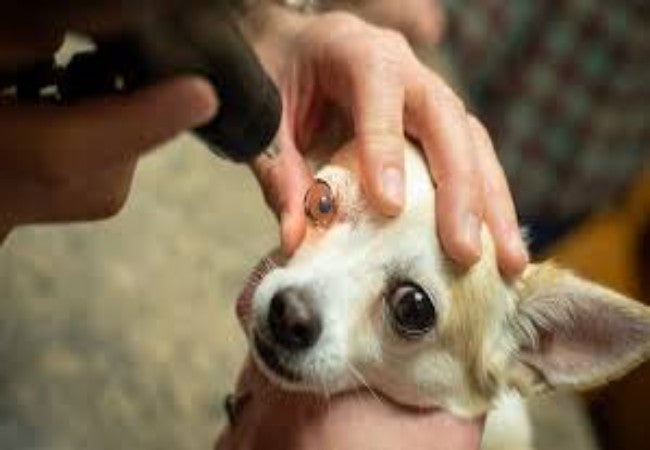
🔬 Diagnostic Approach
- Slit-lamp or ophthalmoscopic exam to identify pattern and layer involvement.
- No systemic disease is associated—contrasts with corneal degeneration.
- Advanced imaging (e.g., corneal OCT) or referral to ophthalmology for endothelial assessment.
- Differentiation from corneal degeneration, ulcer, and infection via history and clinical appearance.
💊 Management & Treatment
- Epithelial/stromal: Typically benign—no treatment unless vision or comfort is affected.
- Endothelial: Hypertonic saline drops may reduce edema; bullae require systemic and topical care.
- Surgical interventions (PTK, keratoplasty or corneal transplants) are considered for painful or vision‑impairing cases.
📈 Prognosis & Monitoring
- Generally excellent for superficial dystrophies—vision unaffected long-term.
- Endothelial forms may recur post-surgery; bullae risk remains.
- Annual ophthalmic recheck to monitor progression and early detection of complications.
🧬 Genetic & Breeding Considerations
- Inherited autosomal patterns; symmetrical presentation supports a genetic cause.
- Breeder screening—exclude affected dogs to reduce the incidence in future litters.
✅ Vet Tips by Dr Duncan Houston BVSc
- 🔍 Include corneal screening in routine eye exams for predisposed breeds.
- 🙂 Educate owners—most dystrophies are harmless; reassure when mild.
- 🚨 Act promptly on signs of ulcers or blisters—ultrasound or referral for advanced care may be needed.
- 📆 Monitor progression yearly and update care as needed.
If your dog's eyes show gray or crystal-like spots or cloudiness—especially in breeds like Shelties, Cavaliers, or Beagles—book an eye check through AskAVet.com. Early detection ensures comfort and clear vision lasting years. 🐾❤️


