Vet Guide to Dislocated Hip in Dogs 2025 🐶🩺

In this article
Vet Guide to Dislocated Hip in Dogs 2025 🐶🩺
By Dr. Duncan Houston BVSc
Dislocated hip, or coxofemoral luxation, occurs when the femoral head (ball) pops out of the acetabulum (socket), disrupting joint capsule, ligaments, muscles, and bones. This usually happens after trauma—like being hit by a car—or in dogs with predisposing conditions such as hip dysplasia, osteoarthritis, or Legg‑Calvé‑Perthes disease.
⚠️ Signs & Symptoms
- Severe pain and inability to bear weight on the affected hind limb — often carried or non‑weightbearing.
- The affected leg appears shorter and may be rotated inward or outward, with swelling and warmth around the hip.
- Reluctance to walk, jump, or stand; reduced appetite from discomfort.
🔬 Diagnosis
- X‑rays: Confirm luxation, determine direction (craniodorsal most common ~90%), check for fractures or hip dysplasia.
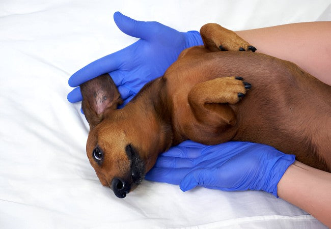
- Physical exam: Palpation may reveal the femoral head displaced; assess for concurrent injuries in trauma cases.
💉 Treatment Options
🔄 Closed Reduction (Non‑Surgical)
- Performed under general anesthesia—joint manually re-seated, then confirmed with X‑ray.
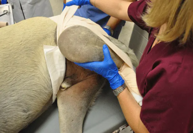
- Apply an Ehmer sling to maintain hip position and restrict movement for 10–14 days, then crate rest for several weeks.
- Success rate ~50%; failure often leads to surgical management.
🔧 Open Reduction (Surgical)
- Required when closed reduction fails or is contraindicated (e.g., fractures, hip dysplasia).
- Techniques include capsulorrhaphy, toggle-pin stabilization, trans-articular pins, iliofemoral sutures; implants remain until healing occurs.
- Success rate ~85–90%, with good restoration of function.
🔪 Alternative Surgical Options
- Femoral head ostectomy (FHO): Removes femoral head, allowing scar tissue to create a functional pseudo-joint; typically restores mobility, no risk of re‑luxation, suitable for chronic or complicated cases.
- Total hip replacement: Artificial joint replacement for severe hip pathology not amenable to other repairs.
🛡️ Post‑Treatment & Rehabilitation
- Strict rest and sling/crate confinement for 6–8 weeks.
- Pain management and anti-inflammatories as prescribed.
- Gradual reintroduction of physical therapy—starting with passive range-of-motion and controlled walking.
- Monitor for complications: re‑luxation, infection, implant issues, nerve damage, or sling sores.
📈 Prognosis
- Good recovery is expected with early and appropriate treatment. Closed reduction success ~50%, open reduction ~85–90%.
- FHO offers solid long-term function, especially in small dogs or severe joint damage.
- Rehab and controlled activity post‑recovery are vital for optimal gait and muscle strength.
✅ Dr Houston’s Clinical Tips
- 🚨 Act fast—closed reduction is more effective within 48 hours.
- 🩺 Confirm reduction via X‑ray and apply Ehmer sling properly; recheck often.
- 🏥 Choose surgery if underlying conditions exist or if closed reduction failed.
- 🛋️ Emphasize scheduled confinement and structured rehab to prevent relapse.
- 📱 Connect via AskAVet.com for recovery guidance or if signs of re‑luxation appear (limping, pain, swelling).


