

Vet Guide to Distichiasis in Dogs 2025 🐶🩺
In this article
Vet Guide to Distichiasis in Dogs 2025–🐶🩺
By Dr. Duncan Houston BVSc
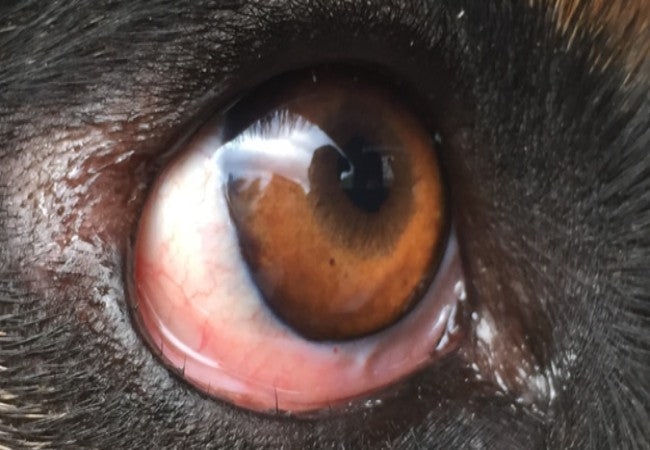
Distichiasis refers to extra eyelashes emerging from the meibomian gland openings along the lid margin—abnormal growth that may brush the cornea and cause damage 😣.
📍 Causes & Breeds
- Congenital, inherited defect—exact genetic mechanism unknown.
- Common breeds include Cocker Spaniels, Cavalier King Charles Spaniels, Bulldogs, Dachshunds, Shih‑Tzus, Poodles, Flat‑Coated Retrievers, Parson Russells, Boxers, Boston Terriers & Yorkies.
⚠️ Clinical Signs
- Often an incidental finding in asymptomatic dogs.
- When lashes irritate: chronic tearing (epiphora), blepharospasm (squinting), conjunctival redness, corneal ulcers, scarring & discomfort.
🔬 Diagnosis
- Detailed eyelid examination, often using magnification or a slit-lamp to detect abnormal lashes.
- Fluorescein staining checks for corneal damage; tear production is assessed with the Schirmer test.
- Differentiation needed: ectopic cilia (follicles through the conjunctiva) are often more painful and ulcerogenic.
💊 Treatment Options
- Benign neglect: No treatment if lashes are soft and asymptomatic.
- Lubricants: Artificial tears to reduce irritation.
- Manual epilation: Plucking with forceps every 4–6 weeks—temporary relief.
- Cryotherapy: Freezing follicles with nitrous oxide or liquid nitrogen; repeat freeze‑thaw cycles, 85–90 % effective.
- Electroepilation or electrocautery: Destroys the follicle with an electric current—localized option.
- Surgical excision: Removal of follicle-bearing area in select cases; requires anesthesia and ophthalmologist referral.
🛡️ Prognosis & Follow‑Up
- Treated dogs generally recover well; new hairs may emerge, especially in young dogs or Shih‑Tzus.
- Ongoing monitoring: watch for recurrence, monitor corneal health, and tear function.
- Risks: Cryo/electro treatments may cause swelling, depigmentation, or scarring; repeated sessions may be needed.
✅ Dr Houston’s Clinical Tips
- 🔍 Always check eyelid margins in breeds predisposed to epiphora.
- 📸 Magnification is essential—asymptomatic dogs are often overlooked until corneal damage.
- 🍃 Use lubricants and epilation for mild cases; escalate to cryo/electro only when needed.
- 📅 Schedule re‑checks post‑procedure at 4–6 weeks and periodically thereafter.
- 🛠️ Refer to a veterinary ophthalmologist for surgical or complex cases.
If your dog is tearing excessively, squinting, or rubbing their eyes, request a thorough ophthalmic exam—even if eyelashes seem normal. Early diagnosis and targeted treatment help prevent corneal ulcers and preserve vision. 🐾❤️


