

Vet’s 2025 Guide to Canine Leishmaniasis Visceral & Cutaneous Care🩺
In this article
Vet’s 2025 Guide to Canine Leishmaniasis Visceral & Cutaneous Care🩺
By Dr. Duncan Houston BVSc
💡 Introduction
Canine leishmaniasis is a vector-borne disease caused by Leishmania parasites, primarily L. infantum. It includes visceral and cutaneous forms, affecting internal organs or skin. Dogs are both victims and important reservoirs, implicating human health in endemic regions. In 2025, veterinarians will use advanced diagnostics, combination therapies, and updated prevention strategies. This guide covers causes, signs, testing, treatments, prevention, and zoonotic risk—plus support from Ask A Vet. 🐾
1. What Is Canine Leishmaniasis?
A disease caused by protozoal parasites transmitted through the bite of infected female phlebotomine sand flies. Dogs infected with L. infantum can remain asymptomatic for months or years before showing signs.
1.1 Forms of Leishmaniasis
- Visceral (“black fever”): affects organs—weight loss, fever, enlarged lymph nodes/spleen, nosebleeds, diarrhea.
- Cutaneous: causes skin ulcers, alopecia, scaling, blepharitis, and interdigital ulcers.
- Mucocutaneous: lesions affecting mucous membranes (rare in dogs).
2. How Is It Transmitted?
Primarily transmitted via sandfly bites. In some areas (like North American Foxhounds), direct dog-to-dog transmission—blood, secretions, or congenital—has been documented.
3. Which Dogs Are at Risk?
- Dogs in endemic regions—Mediterranean, Latin America, Middle East, parts of Africa & Asia.
- Imported dogs from these regions—cases reported in the U.S. and Canada.
- No strong breed predilection, though some groups (Foxhounds, Neapolitan Mastiffs, Spinone) show higher infection rates.
4. Clinical Signs to Watch
Signs vary widely—regular screening is essential in at-risk dogs.
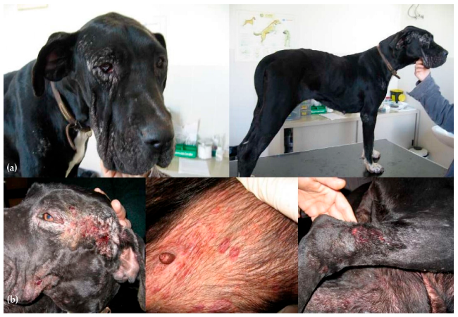
4.1 Cutaneous signs:
- Alopecia, scaling, exfoliative dermatitis
- Ulcers on ears, footpads, muzzle (see Figures above).
- Nail overgrowth (onychogryphosis)
4.2 Visceral signs:
- Weight loss, poor appetite, lethargy
- Diarrhea, vomiting, tarry stools.
- Epistaxis (nose bleeds), swollen lymph nodes
- Kidney failure signs—increased drinking & urination.
- Ocular signs: blepharitis, conjunctivitis
5. Diagnosing Leishmaniasis in 2025 🧪
Combine clinical exam with definitive lab testing.
5.1 Serological testing
- ELISA or immunofluorescence to detect antibodies.
5.2 PCR & Cytology
- PCR on blood or aspirates is highly specific and confirms active infection.
- Fine-needle aspirates—detect amastigotes in lymph nodes, skin lesions.
5.3 Staging & Organ Evaluation
- Chest X-rays/abdominal ultrasound for organ involvement
- Bloodwork: CBC, biochemistry, kidney function tests (BUN, creatinine)
- Urinalysis with protein-to-creatinine ratio—identifies kidney damage.
A standardized staging system from LeishVet guides prognosis and therapy.
6. 2025 Treatment Approaches ❤️
No cure—but treatment suppresses infection, resolves signs, and prolongs life.
6.1 Drug combinations
- Allopurinol + Meglumine antimoniate
- Allopurinol + Miltefosine
- Amphotericin B is reserved for resistant or renal cases.
6.2 Monitoring & Management
- Regular bloodwork and renal monitoring
- Re-check parasite load via serology/PCR
- Co-manage kidney and ocular complications
Treatment can be lifelong; relapse is common—follow-ups every 6–12 months are advised.
6.3 Emerging Vaccine Protection
Live-attenuated vaccines (LmCen–/–) show early promise in immunizing dogs against visceral disease.
Commercial vaccines in Brazil and Europe (Leishmune, CaniLeish, Letifend) are used in endemic regions—with efficacy of 70–87%.
7. Prevention Strategies 🔒
- Insecticide-impregnated collars (deltamethrin) reduce risk by ~86%.
- Spot-on repellents paired with collars are common in Europe.
- Keep dogs indoors during peak sandfly activity (dusk–dawn)
- Environmental control: remove breeding sites and deploy bed nets.
- Vaccination: best in endemic areas, used alongside vector control.
8. Zoonotic Considerations 🌍
Though L. infantum is zoonotic, natural transmission from dogs to humans is rare when vector control is in place. However, managing canine reservoirs is essential to reduce risk.
9. How Ask A Vet Helps 🩺
- Provide reminders for treatment and vaccinations
- Support monitoring: track appetite, skin changes, and weight
- Photo-based symptom tracking of lesions or swelling
- Home guidance on collared care, environment, and when to visit the vet
10. Prognosis & Follow-Up
- Visceral cases: Carefully managed dogs can live several years with improved quality of life.
- Cutaneous-only cases: Good prognosis with localized therapy and vector control.
- Relapses occur in ~20–30% of cases, so lifelong monitoring is key.
🔍 Summary Takeaways
- Leishmaniasis is complex and systemic—watch for skin, gland, and organ signs
- Diagnosis uses blood tests, PCR, imaging, and staging systems
- Treatment combines antiparasitic drugs with ongoing medical supervision
- Prevention via collars, repellents, indoor care, and novel vaccines is essential
- Minimizing canine infection helps protect human health
- Ask A Vet enhances care through reminders, monitoring, and support
Conclusion ❤️
In 2025, managing canine leishmaniasis requires thorough diagnostics, strategic treatment, and comprehensive prevention—especially in endemic or travel-prone dogs. While it remains a life-affecting disease, early detection and consistent care can significantly improve outlooks for dogs and public health. Ask A Vet offers the ongoing support you need to help your dog live well in every stage. 🐕✨
Dr Duncan Houston BVSc – your guide to responsible, compassionate parasitic disease management.
Learn more at AskAVet.com and download the Ask A Vet app for personalized support, reminders, and vet access any time. ❤️


