Vet’s 2025 Guide to Canine Papilledema (Optic Disc Swelling) 🧠✨🩺

In this article
Vet’s 2025 Guide to Canine Papilledema (Optic Disc Swelling) 🧠✨🩺
By Dr. Duncan Houston BVSc
💡 What Is Papilledema?
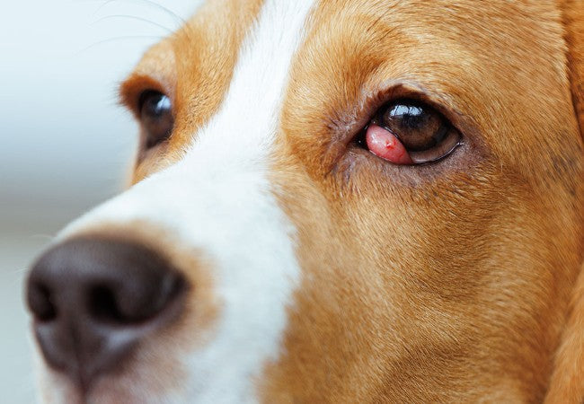
Papilledema refers to swelling of the optic disc due to increased intracranial pressure (ICP), causing the optic nerve head to bulge into the eye. It's a critical 🔴 sign that vision and brain function may be at risk.
🚩 Why It Matters
- ⏱️ Signals serious neurologic disease—needs prompt veterinary care.
- ⚠️ May cause vision loss if elevated ICP remains untreated.
- 🧬 Reflects disorders like tumors, hydrocephalus, inflammation, trauma, or venous blockage.
👀 Who Is Affected?
- Dogs with intracranial masses—up to 50% show papilledema on fundus exam.
- Cases involving hydrocephalus, meningitis, encephalitis, head trauma or cerebral edema.
- Both optic nerves are typically affected, but unilateral papilledema may signal a local lesion like optic glioma.
🔍 Clinical Signs
- 👁️ Raised optic disc with blurred margins, peripapillary hemorrhage, and venous congestion.
- 👀 May see enlarged blind spots or early visual field loss.
- 🔦 Pupils often normal early—later signs like vision loss or fixed pupils if severe.
- 🐾 Neurologic deficits may accompany: behavior change, ataxia, seizure, appetite loss, lethargy.
🧪 Diagnosing Papilledema
- Ophthalmoscopy / Fundus Photography: reveals optic disc elevation, blurred margins, venous swelling, peripapillary hemorrhages.
- Optical coherence tomography (OCT): advanced imaging detects disc swelling, subclinical changes.
- Ultrasound/OCT sheath diameter can indirectly suggest raised ICP in emergencies.
- Brain imaging (MRI/CT): essential to identify underlying cause—tumors, hemorrhage, hydrocephalus.
- Neurologic & systemic work-up: CBC, biochemistry, coagulation, infectious disease tests, CSF analysis.
🛠 Treatment Strategies
- 🎯 Treat underlying cause: surgical removal or radiation for tumors; antibiotics/antifungals or steroids for inflammation/infection.
- 💧 Reduce ICP: administer diuretics (acetazolamide, furosemide), occasional serial CSF drainage.
- 🧠 Supportive care: hospitalize for fluid balance, oxygen support, maintain head elevation, anti-seizure meds
- 👁️ Corticosteroids: help reduce inflammation or tumor-associated swelling—use judiciously.
- 🧪 Co-management: manage hypertension, coagulation disorders, or systemic disease.
📈 Prognosis & Monitoring
- 🟢 Good prognosis if the cause is identified and treated early.
- 🟡 Outcomes vary based on disease type/severity; papilledema may resolve in days to weeks if ICP drops.
- 🔴 Without treatment, may result in permanent vision loss or fatal complications like brain herniation.
- 🔁 Risk of recurrence if cause not fully resolved—monitor with serial exams & imaging.
🏡 Ask A Vet App Home‑Monitoring Tools 📲🐶
- 📷 Upload fundus photos or videos for vet review.
- 🔔 Record neurologic/red-flag signs: vision changes, disorientation, seizures.
- 📅 Schedule ICP-reducing medication reminders and recheck imaging.
- 📘 Built-in info: “ICP reduction tips,” “Fundoscopy basics,” “Brain emergency signs.”
🔑 Key Takeaways 🧠✅
- Papilledema signals dangerously high intracranial pressure—urgent treatment needed.
- Causes range from tumors, inflammation, infection, trauma, to venous congestion.
- Diagnosis uses ophthalmoscopy, OCT, neuroimaging & systemic tests.
- Treatment targets the underlying cause, plus ICP control and supportive care.
- Prognosis depends on early intervention; many recover if the disease is managed promptly.
- Ask A Vet tools support monitoring, vet communication, red‑flag alerts & treatment adherence.
🩺 Final Thoughts ❤️
In 2025, veterinary care for papilledema combines precise ocular diagnosis, effective ICP management, and cause-focused treatment, all supported by proactive owner monitoring. With tools like Ask A Vet, owners can help catch changes early—whether in behavior, vision, or neuro‑signs—and empower their vet team with fundus images and symptom logs. This partnership can save vision and lives for dogs facing this critical condition 🐾✨.
Visit AskAVet.com and download the Ask A Vet app to upload ocular photos, log symptoms, set meds reminders, receive alerts, and keep your veterinarian updated throughout your dog’s neurological care. 📲🐶


