🩺 Vet’s 2025 Guide to Canine Primary Lens Luxation 🐶 Focused Care for a Painful Eye Condition

In this article
Vet’s 2025 Guide to Canine Primary Lens Luxation 🐶 Diagnosis, Surgery & Ongoing Care
By Dr. Duncan Houston BVSc
💡 Introduction
Primary lens luxation (PLL) is a hereditary condition where the zonular fibers that hold the lens weaken and break, causing displacement of the lens, either forward (anterior) or backward (posterior). This leads to a painful eye, inflammation, and a high risk of secondary glaucoma. Early detection and intervention are key to preserving vision and eye comfort in affected dogs.
1. Understanding PLL 🧠
- Genetic inheritance: PLL follows an autosomal recessive pattern, most commonly due to mutations in the ADAMTS17 gene.
- Breed predispositions: Seen in many terrier breeds (Jack Russell, Fox, Yorkshire, West Highland White), Australian Cattle Dogs, Shar-Pei, Chinese Crested, Border Collie, American Eskimo, among others.
- Age of onset: Typically between 3–8 years old; often unilateral initially, but the second eye is frequently affected later.
2. Clinical Signs & Emergency Cues 🚨
- Painful, red eye: intense tearing, squinting, and blepharospasm.
- Cloudy cornea: may display an aphakic crescent sign if subluxated.
- Increased eye pressure: elevated IOP signals glaucoma risk, especially with anterior luxation.
- Pain from corneal touch: lens forward presses against cornea, causing edema and significant discomfort.
- Vision loss: potential if untreated, especially with anterior luxation and secondary glaucoma.
3. Diagnostic Work‑up in 2025 🧪
- Complete ophthalmic exam: slit-lamp, ophthalmoscope, tonometry, Schirmer tear test.
- Ultrasound: aids assessment when cornea is cloudy.
- Genetic testing: confirm carrier/affected status with ADAMTS17 mutation analysis.
- Evaluate fellow eye: frequent exams for early signs; prophylactic miotics like demecarium may delay second-eye luxation.
4. Urgency: Anterior vs Posterior Luxation
- Anterior luxation: medical emergency due to glaucoma and corneal damage—requires prompt surgical removal.
- Posterior luxation: less urgent; pupils can be constricted to prevent forward movement, with close monitoring.
5. Treatment Options & Protocols ❤️
5.1 Surgical Removal (Lensectomy)
- Intracapsular extraction: performed by a veterinary ophthalmologist; quick removal of lens.
- Enucleation: considered if the eye is blind, painful, or if glaucoma cannot be controlled.
- Risks & prognosis: guarded—possible postoperative glaucoma, retinal detachment, uveitis.
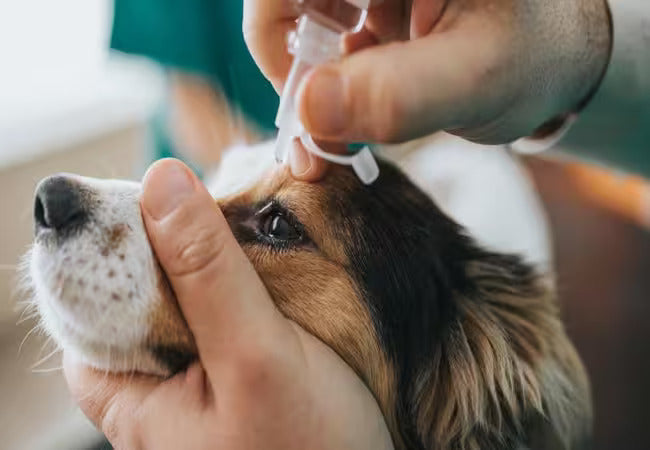
- Post-op care: strict rest; hospital stay 1–3 days; topical/systemic anti‑inflammatories and glaucoma medications; cone use for 2 weeks.
5.2 Medical Management (Conservative)
- Miotic therapy: demecarium or similar agents to constrict pupil and prevent forward lens movement; suitable for mild or posterior cases.
- Glaucoma/uveitis control: pain relief, pressure-reducing drops, corticosteroids.
- Monitoring: regular IOP checks, vision testing, and exams every 1–3 months.
6. Prognosis & Long-Term Monitoring
- Vision preservation: with early surgical intervention in anterior luxation, ~70–80% retain vision; posterior cases often stable conservatively.
- Complications: glaucoma, retinal detachment, and chronic uveitis; all require lifelong surveillance.
- Prophylaxis: miotic therapy on the healthy eye may delay luxation onset.
7. Breeding Considerations & Prevention 🔬
- DNA testing: screen breeding stock for ADAMTS17 mutation; avoid breeding carriers or affected dogs.
- Early checkups: biannual eye exams from age 2–3 in at-risk breeds.
- Client education: ensure owners know the signs of PLL and glaucoma emergencies.
8. Ask A Vet Home Support 🏡
- Symptom tracking: eye redness, squinting, tearing, vision changes via app.
- Medication reminders: miotics, anti-inflammatories, IOP drops.
- Photo/video submissions: pupil position, discharge, pain behaviors.
- Recovery guides: post-lensectomy rest reminders, cone use, leash-only walks.
- Follow-up alerts: schedule annual eye exams, IOP monitoring.
🔍 Key Takeaways
- PLL is an inherited, painful, sight-threatening disease common in terriers and several breed lines.
- Anterior luxation is an emergency—requires prompt surgical removal to preserve vision.
- Posterior luxation may be managed medically, but vision and glaucoma risks persist.
- Lifelong monitoring post-treatment is essential due to potential complications.
- Genetic testing prevents breeding of carriers; regular exams aid early detection.
- Ask A Vet empowers owners with real-time monitoring and recovery support.
🩺 Conclusion ❤️
Primary lens luxation is a significant hereditary eye disease that demands early recognition and decisive intervention. In 2025, advanced diagnostics, genetic screening, precise surgery, and dedicated home monitoring via Ask A Vet enable optimized outcomes—even in breeds at high risk. With informed care and vigilant support, many dogs can retain sight and comfort through their golden years. 🐶✨
Dr Duncan Houston BVSc – advocating for vision health, surgical precision, and owner-led care.
Visit AskAVet.com and download the Ask A Vet app for eye health reminders, symptom tracking, photo uploads, and specialist guidance through every step of lens luxation care. ❤️


