

Vet’s 2025 Guide to Canine Retinal Detachment 🩺 Detection, Treatment & Prevention
In this article
Vet’s 2025 Guide to Canine Retinal Detachment 🩺 Detection, Treatment & Prevention
By Dr. Duncan Houston BVSc
💡 What Is Retinal Detachment?
Retinal detachment is the separation of the retina from the underlying tissue, leading to visual impairment or blindness. It can be rhegmatogenous (tear-related), exudative (fluid accumulation), or tractional (scar or membrane pulls).
On fundus exam, the retina may appear as a grey “veil” with visible blood vessels that seem to float anteriorly.
🚩 Who’s Affected & Why?
- Any breed or age—but more common in older dogs, or those with congenital defects like Collie Eye Anomaly or retinal dysplasia.
- Underlying contributors: systemic hypertension, uveitis/chorioretinitis, glaucoma, ocular tumors, trauma, or metabolic/fungal disease.
- Genetic predispositions—breeds with inherited eye conditions should be screened and not bred.
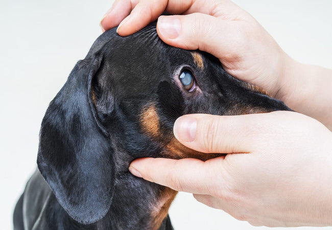
👀 Clinical Signs
- Sudden vision loss—bumping into objects, failing to catch toys.
- Dilated pupil(s), absent or delayed pupillary reflexes.
- Visible retina vessels and a grey membrane on ophthalmoscopy.
- Partial detachments may preserve some vision; complete detachments usually result in blindness.
- May accompany uveitis, glaucoma, or intraocular hemorrhage.
🧪 Diagnostic Tools
1. Ophthalmoscopy
Direct or indirect exam reveals detached retina—vessels in different focus planes, paleness, grey sheen.
2. Ultrasonography
Used when ocular opacity prevents direct view; shows “seagull sign” membrane in vitreous.
3. Systemic Work‑Up
- Measure blood pressure to detect hypertension.
- Run CBC, chemistry, urinalysis; include tests for uveitis-related diseases.
- Screen for infectious or neoplastic causes (fungal, rickettsial, systemic neoplasia).
- Advanced imaging (OCT, electroretinography) if needed.
🩺 Treatment Approaches
1. Address Underlying Causes
- Control hypertension—often results in spontaneous resolution of exudative detachments.
- Treat uveitis or infections with appropriate therapy.
- Manage glaucoma, tumors, or systemic illness to prevent progression.
2. Surgical Repair
Referral to veterinary ophthalmologist recommended. Options include:
- **Barrier retinopexy** via diode laser for small tears.
- **Vitrectomy**: removal of vitreous and reattachment with gas or silicone oil.
- **Scleral buckling**: silicone band to stabilize retina.
- **Cryoretinopexy/laser** seal tears in conjunction with other methods.
Prompt treatment offers better chance of vision recovery, though full restoration isn't guaranteed.
3. Medical Management & Follow‑Up
- Anti‑inflammatories (topical/systemic) for uveitis.
- IOP-lowering meds if glaucoma is present.
- Regular ophthalmic exams to monitor reattachment or complications (glaucoma, cataracts, uveitis).
📈 Prognosis & Complications
- Exudative detachments from systemic disease may resolve with medical control.
- Surgical detachments have variable outcomes; macula involvement reduces visual prognosis.
- Untreated cases may lead to permanent detachment, chronic uveitis, secondary glaucoma, or eye pain.
- Early detection and referral improve chances for useful vision.
🚫 Prevention Strategies
- Monitor at-risk breeds with frequent ophthalmic exams.
- Control risk factors—manage blood pressure, treat uveitis/infection/glaucoma early.
- Avoid trauma; handle eye conditions promptly.
🏡 Ask A Vet Home Support
- 🗓 Set reminders for blood pressure checks, medication, follow‑ups.
- 📸 Upload videos of movements or behavior suggesting vision loss.
- 📊 Log symptoms—bumping, pupil size, light reflex changes.
- 🔔 Alerts for vision loss, eye redness, pain, or cloudy eyes.
- 📚 Guides: home eye exams, recognizing vision impairment, post-surgical care.
🔑 Key Takeaways
- Retinal detachment is a vision-threatening emergency—look for sudden blindness, dilated pupils, and retina veil on exam.
- Diagnose with fundus exam and ultrasound; work-up underlying disease.
- Treatment depends on cause; surgery may restore vision if timely.
- Regular monitoring prevents complications like glaucoma or chronic inflammation.
- Ask A Vet helps with home monitoring, reminders, guidance, and early alerts.
🩺 Final Thoughts ❤️
In 2025, with modern ocular imaging, laser and surgical repair, and integrated care via the Ask A Vet app, dogs with retinal detachment have improved chances for vision recovery. Prompt detection, early treatment, and involved owner support make all the difference in preserving sight and quality of life. 🐾✨
Visit AskAVet.com and download the Ask A Vet app to track vision signs, upload videos, schedule medication and exam reminders, and stay connected with your veterinary ophthalmologist—directly from your phone. 📲


