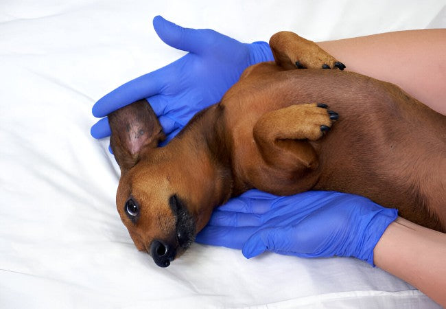
Vet’s 2025 Guide to Canine Salivary Mucocele (Sialocele) 🐶✨🩺
In this article
Vet’s 2025 Guide to Canine Salivary Mucocele 🩺 Diagnosis, Treatment & Recovery
By Dr. Duncan Houston BVSc
💡 What Is a Salivary Mucocele?
A salivary mucocele, or sialocele, is a mucus-filled swelling caused by leakage of saliva from a damaged gland or duct into surrounding tissues, forming a soft, fluctuant, and usually painless cavity.
🚩 Why It Matters in 2025
- 📈 With accessible imaging (CT, ultrasound), veterinarians can define location and plan precise surgeries.
- 📏 Timely identification reduces risks of dysfunction—breathing/swallowing compromise in pharyngeal mucocele.
- 🤝 Owners can now use remote monitoring tools like the Ask A Vet app to log swelling changes and symptoms in real-time.
🧬 Where Do They Occur & Types
Sialoceles are classified by gland or duct location—each causes distinctive swelling:
- Cervical (mandibular + sublingual glands): most common—lump under jaw/neck.
- Sublingual (“ranula”): swelling under tongue—may bleed or obstruct eating.
- Pharyngeal: deeper swelling in throat—can impair swallowing and breathing.
- Zygomatic: rare—affects cheek area near the eye.
👀 Who Gets Affected?
- 🦮 Any breed or age can develop sialoceles — but predisposition noted in German Shepherds, Dachshunds, Poodles, Silky Terriers.
- 🔍 Mostly seen in dogs, rarely in cats.
🧠 Causes & Risk Factors
- 🔨 Trauma: leash jerks, bite wounds, foreign bodies—most common.
- 🧱 Sialoliths (salivary stones) can rupture ducts and precipitate mucocele formation.
- 🩻 Iatrogenic injury: post-surgical or post-extraction trauma.
- 🏥 Idiopathic: occasionally cause remains unknown.
- 🎯 Cervical gland pairing often leads to combined mandibular and sublingual gland involvement.
👀 Clinical Signs
- 💧 Soft, fluctuant, non-painful swelling near affected gland—slow developing.
- 🍴 Dysphagia or drooling when swelling impinges the mouth or throat.
- 😷 Respiratory noise or difficulty if pharyngeal mucocele compresses airway.
- 🩸 Bleeding or infection possible, particularly in ranula with trauma.
- ⚠️ Rare cases show osseous metaplasia—hard calcified pseudocapsule.
🔍 Diagnosis
- Physical exam & history: palpation of swelling; note fluctuations, growth, pain.
- Fine‑needle aspiration (FNA): harvest ropy, viscous, yellow or blood‑tinged saliva; low cellularity confirms mucocele.
- Imaging: Ultrasound or CT confirm size, location—CT aids surgical planning in complex cases.
- Rule out other issues: differentiate from abscesses, neoplasia via aspiration cytology, culture, maybe biopsy.
🛠 Treatment Options
1. Surgical Removal (Gold Standard)
- ✂️ Sialoadenectomy: removal of affected gland(s) plus duct—often both mandibular & sublingual if cervical or sublingual mucocele.
- 🛠️ Complex locations (zygomatic/pharyngeal) may require specialist referral.
- 🩺 Surgical complications rare; recurrence <0.5% when performed correctly.
2. Aspiration & Drainage
- 🩹 Temporary relief—fluid drained with needle but mucocele often recurs.
- ⚠️ Risk of infection from repeated aspiration—sterile technique essential.
3. Marsupialization (Sublingual Only)
- 🔄 Create permanent opening allowing ranula to drain orally—may avert full gland removal.
- 🩻 Effectiveness varies; sometimes used with gland removal.
4. Supportive Care
- 💉 Pain management and anti-inflammatories.
- 🩼 Soft foods reduce trauma during healing phase.
- ⚕️ Monitor for infection or hemorrhage post‑drainage.
📅 Follow-up & Prognosis
- 🟢 Excellent prognosis post-surgery; most dogs fully recover.
- 🟡 Drainage alone has high recurrence rate; often delays surgery.
- 🔴 Rare cyst calcification occurs—requires pseudocapsule and gland removal.
- 🕵️♀️ Monitor for regrowth; recurrence very rare after complete excision.
🏡 Ask A Vet App Home‑Monitoring Tools 📲🐶
- 📊 Track lump size and changes in swelling daily.
- 🗓️ Set reminders for aspiration appointments or surgery follow‑up.
- 📷 Upload photos of swelling or surgical site to share with vet.
- 🔔 Alert if breathing, swallowing difficulty or infection signs appear.
- 📘 Guides: “Post‑op care,” “Drain site cleaning,” “Soft food recipes.”
🔑 Key Takeaways 🧠✅
- Sialoceles are common, painless salivary swellings—cervical type most frequent.
- Diagnosis: FNA, imaging, rule out tumors or infection.
- Sialoadenectomy is gold standard; drainage provides temporary relief.
- Excellent prognosis post-surgery; recurrence rare.
- Ask A Vet app helps owners monitor, manage treatment, and alert vets early.
🩺 Final Thoughts ❤️
In 2025, managing canine salivary mucoceles combines accurate diagnosis, expert surgical removal, and supportive aftercare. Timely detection lessens the risk of complications like airway obstruction. With tools like the Ask A Vet app, owners stay connected—logging swelling, uploading images, and keeping track of post-op care. This team-based approach ensures dogs recover fully and stay comfortable, with minimal recurrence 🐾✨.
Visit AskAVet.com and download the Ask A Vet app to log lump changes, schedule treatments or surgery reminders, upload photos, receive alerts, and stay connected with your vet throughout your dog’s recovery. 📲🐶


