

Vet’s 2025 Guide to Canine Vesiculopustular Dermatoses 🩺 Blisters, Pustules & Treatment Tips
In this article
Vet’s 2025 Guide to Canine Vesiculopustular Dermatoses 🩺 Blisters, Pustules & Treatment Tips
By Dr. Duncan Houston BVSc
💡 What Are Vesiculopustular Dermatoses?
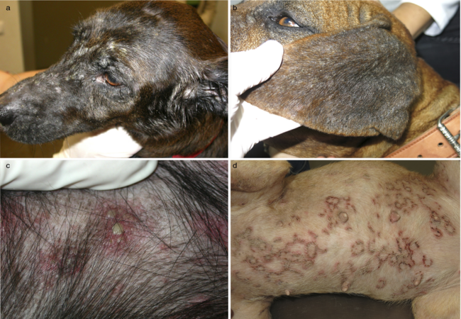
Vesiculopustular dermatoses refer to a group of skin conditions characterized by the presence of vesicles (clear, fluid-filled blisters) and/or pustules (pus-filled bumps) on the skin of dogs 🐶. These lesions signal inflammation and can result from infections, allergies, autoimmune disorders, or genetic skin diseases.
🚩 Causes & Risk Factors
- 🦠Infections (common):
•Superficial pyoderma – bacterial infection causing pustules.
•Dermatophytosis (ringworm) – rare, causes vesicles/pustules in young dogs.
•Demodex (mange) – mite infestations with pustular lesions. - ⚠️Allergic or reactive:
• Sterile eosinophilic pustulosis — allergic reaction, common in terraces.
• Drug eruptions – adverse reactions to medications. - 🧬Autoimmune blistering disorders:
• Pemphigus foliaceus — pustular lesions starting on face/pads.
• Pemphigus vulgaris — more severe, ulcers at mucocutaneous junctions.
• Bullous pemphigoid — tense blisters on skin and mucosa.
• Linear IgA dermatosis — Dachshunds cause pustules beneath skin.
• Subcorneal pustular dermatosis — sterile pustules, often Schnauzers.
• Lupus erythematosus (SLE, DLE) – vesicles and crusting lupus conditions.
• Dermatomyositis – collies and Shelties with pustular lesions.
👀 Recognizing the Clinical Signs
Dogs with vesiculopustular skin issues may show:
- Clear or cloudy fluid-filled blisters (vesicles)
- Pustules—raised bumps with pus
- Scabs, crusts and scaling from ruptured lesions
- Hair loss and redness (erythema)
- Blisters in flexural areas (armpits, groin), footpads, nose, muzzle, anus/urogenital junction
- Possible itching, pain, licking, secondary bacterial odour/infection
- Systemic signs (rare): lethargy, fever, joint pain in severe autoimmune cases
🧪 Diagnosis: A Stepwise Approach
- Thorough history: onset, pattern, breed predisposition, medications, previous disease.
- Physical exam: full-body skin check, look for lesion distribution, mucosal involvement.
- Skin cytology: impression smears or pustule contents to check bacteria, yeast, eosinophils.
- Skin scraping: rule out demodex, mites.
- Fungal culture: test for ringworm.
- Blood tests: CBC, chemistry, urinalysis to assess for systemic disease like lupus.
- Skin biopsy & histopathology: gold standard for autoimmune diseases (pemphigus, lupus, pemphigoid). Biopsy intact lesions.
- Immunofluorescence/ELISA: identify autoantibodies (e.g., BP180, BP230 in bullous pemphigoid).
🛠 Treatment Strategies by Cause
1. Infection-based Therapies
- Antibiotics: oral/topical for pyoderma (first-tier: cephalosporins; resistant cases: non-beta-lactams).
- Antifungals: for confirmed dermatophytosis (terbinafine, itraconazole).
- Parasiticides: treat demodex (topical or systemic).
- 🏥 Supportive care: medicated baths, cleanse lesions to reduce odors and prevent infections.
2. Autoimmune and Sterile Conditions
- Glucocorticoids: systemic prednisone/prednisolone as primary immunosuppressive therapy.
- Steroid-sparing agents: azathioprine, mycophenolate, chlorambucil, cyclosporine, methotrexate—tailored per disease.
- Drug-specific therapies: dapsone for subcorneal pustular dermatosis or linear IgA, tetracycline+niacinamide for pemphigus conditions.
- Topical therapy: potent corticosteroids or tacrolimus for localized lesions.
- Sun protection: photosensitive autoimmune diseases need UV avoidance.
3. Supportive & Adjunct Care
- 🛁 Regular antimicrobial baths (chlorhexidine, benzoyl peroxide) to support lesion healing.
- 🐾 Gentle wound management: cleanse, ointments, prevent licking with e-collar.
- 💧 Nutritional support: omega-3 fatty acids, vitamin E for skin barrier health.
- 🩺 Pain relief: NSAIDs or opioids if lesions are painful.
- 🏥 Inpatient care for severe cases (SLE, pemphigus vulgaris, bullous pemphigoid).
📈 Monitoring, Prognosis & Recovery
- 🩺 Follow-up visits every 2 weeks initially—skin checks, blood tests for meds side effects.
- 📉 Medication tapering once lesions improve, with monitoring for recurrence.
- 🔁 Many autoimmune conditions relapse; lifelong therapy often required for chronic diseases.
- ✅ Infection-based cases have good outcomes with proper treatment.
- ⚠️ Autoimmune prognosis varies: pemphigus foliaceus is often manageable; pemphigus vulgaris and bullous pemphigoid can be serious.
- 🌞 Prevent UV exposure in UV-triggered diseases to avoid flare-ups.
🏡 Ask A Vet App Home‑Support Tools 📱🐾
- 📆 Reminders for meds, baths, biopsy/follow-up appointments.
- 📸 Upload lesion photos for remote vet monitoring.
- 📊 Log lesion count, itchiness, systemic signs.
- 🔔 Alerts for new blisters, crusting or signs of infection.
- 📚 Access care guides: bath protocols, wound care steps, meds dosages.
🔑 Key Takeaways 🧠✅
- 🔄 Vesiculopustular dermatoses range from mild infections to severe autoimmune diseases.
- 🔬 Diagnosis relies on cytology, scraping, biopsy & advanced tests.
- 🩺 Treatment varies from antibiotics/antifungals to powerful immunosuppressives.
- 🛁 Supportive care and sun protection are vital to healing.
- ⚕️ Monitoring for relapses and side effects essential; App tools keep you on track.
🩺 Final Thoughts ❤️🐶
In 2025, canine vesiculopustular dermatoses remain a diagnostic and therapeutic challenge—but with systematic evaluation, targeted therapy, and strong home support via tools like the Ask A Vet app, most dogs can heal fully or manage chronic conditions effectively. Your vigilance, paired with veterinary partnership, is key to your pup’s comfort and recovery. 🐾✨
Visit AskAVet.com and download the Ask A Vet app to schedule meds, log lesions, upload images, set alerts, and stay connected with your vet—anytime, anywhere. 📲🐾


