Veterinary Guide to Canine Bile Peritonitis 2025 🩺🐶

In this article
Veterinary Guide to Canine Bile Peritonitis 2025 🩺🐶
By Dr. Duncan Houston BVSc
🧬 What Is Bile Peritonitis?
Bile peritonitis occurs when bile—normally contained within the gallbladder or biliary ducts—leaks into the abdominal cavity, causing a toxic, inflammatory reaction known as chemical peritonitis. When bacterial contamination occurs, it becomes septic and life-threatening.
👥 Causes & Risk Factors
- Gallbladder mucocele: Mucocele development can stretch and rupture the gallbladder wall.
- Cholecystitis/choledochitis: Necrotizing inflammation weakens ducts/gallbladder.
- Trauma: Blunt abdominal trauma leading to bile duct rupture.
- Neoplasia or choleliths: Tumors or stones obstructing or rupturing bile ducts.
- Gastric or duodenal perforation: Rare causes, like bilious reflux-related leaks.
👀 Clinical Signs
- Early signs are mild: inappetence, vague abdominal discomfort, lethargy.
- Progression to acute pain, distended abdomen, vomiting, diarrhea.
- Systemic signs: fever or hypothermia (septic), pale mucous membranes, tachycardia, weak pulse.
- Jaundice (icterus), yellow urine/stools indicating bile contamination.
- Shock and collapse in severe septic bile leaks.
🔬 Diagnostic Workup
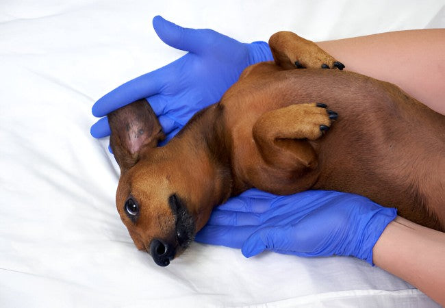
- Physical & history: Focused abdominal palpation, assessment of toxin exposure or trauma.
- Bloodwork: CBC (neutrophilia, left shift), chemistry (elevated liver enzymes, hyperbilirubinemia), urinalysis (bilirubinuria).
- Imaging: Abdominal US ideal—shows effusion, gallbladder abnormalities, duct rupture; X-ray supportive.
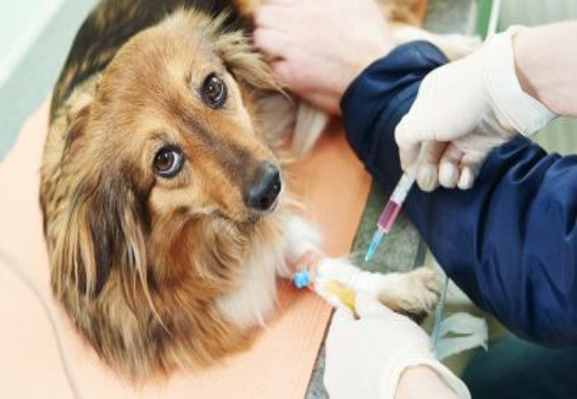
- Abdominocentesis: Fluid analysis reveals gross green/brown bile; cytology with neutrophils and bile pigments.
- Bilirubin assay: Peritoneal fluid to serum bilirubin ratio >2:1 confirms bile leakage.
- Cultures & biopsies: Aerobic/anaerobic culture of fluid and tissue, liver biopsy for gallbladder mucocele or infection.
🚨 Treatment Approach
🏥 Stabilization & Supportive Care
- IV fluids and support for shock, pain control, and electrolyte management.
- Vitamin K if prolonged clotting times, and fresh frozen plasma if bleeding risk.
- Anti-nausea and gastric protectants (antiemetics, H₂ antagonists).
⚙️ Surgical Intervention
- Exploratory laparotomy: Identify and repair the source (CBD, gallbladder).
- Gallbladder removal (cholecystectomy) or duct repair/anastomosis/cholecystoduodenostomy based on findings.
- Thorough abdominal lavage (1–2 L sterile saline ± antibiotics); drains may be placed.
- Liver biopsy and peritoneal fluid sampling for culture and histopathology.
💊 Post-Op & Medical Therapy
- Broad-spectrum antibiotics against Gram-negative and anaerobic bacteria for 4–8 weeks.
- Supportive medications: antiemetics, analgesics, hepatoprotectants (ursodiol, SAMe), probiotics.
- Monitor and correct ileus, shock, and septic signs with ICU care.
📈 Prognosis & Monitoring
- Sterile bile peritonitis: good prognosis; lower mortality if treated quickly.
- Septic bile peritonitis: worse prognosis—sterile vs septic shows 100% vs ~27% short-term survival.
- Recent study: traumatic bile peritonitis—88% survival to discharge after surgery.
- Long-term follow-up: periodic blood testing, imaging for recurrence, and monitoring liver health.
🏠 Home Care & Prevention
- Strict rest 10–14 days post-op; pain medication and feeding per vet instructions.
- Prevent gallbladder issues: manage endocrine disease, maintain healthy weight, monitor predisposed breeds (e.g. Schnauzers, Cocker Spaniels).
- Avoid trauma; schedule regular abdominal ultrasounds in at-risk dogs.
- Strict monitoring for vomiting, abdominal pain, appetite changes.
📱 Ask A Vet Telehealth Support
- 📷 Owners upload incision site and belly photos for remote checking of swelling or discomfort.
- 🔔 Medication and fluid reminders: antibiotics, ursodiol, analgesics, suture/drain care.
- 📅 Follow-up scheduling: automated lab and imaging alerts (CBC, liver panel, abdominal US).
🎓 Case Spotlight: “Milo” the Mini Schnauzer
Milo, a 9‑year‑old Miniature Schnauzer, presented with acute vomiting and jaundice. Ultrasound revealed a gallbladder mucocele and free abdominal fluid. After stabilization, surgery included cholecystectomy and lavage. Cytology confirmed sterile bile peritonitis. He recovered uneventfully and returned home on antibiotics, ursodiol, and low-fat diet. Ask A Vet reminders helped with medications and follow-ups; at 6‑month recheck, he remained healthy and active 🌟.
🔚 Key Takeaways
- Bile peritonitis is a surgical emergency caused by bile leakage, which can be septic or sterile.
- Early diagnosis via imaging and cytology improves outcomes.
- Prompt surgery with lavage and repair + plus medical support, is essential for survival.
- Prognosis depends on sterility of bile—sterile = good, septic = guarded, trauma-treated = favorable.
- Ask A Vet telehealth supports every step—from recovery care to long-term monitoring and preventative advice 📱🐾
Dr Duncan Houston BVSc, founder of Ask A Vet. Download the Ask A Vet app today to guide your dog’s bile peritonitis journey—from emergency to recovery, diet planning, and ongoing wellness 🌟


