

Veterinary Guide to Canine Intervertebral Disc Disease 2025 🐶
In this article
Veterinary Guide to Canine Intervertebral Disc Disease 2025 🩺🐶
By Dr. Duncan Houston BVSc
🧬 What Is IVDD?
Intervertebral Disc Disease (IVDD) occurs when spinal discs degenerate, bulge, or herniate, compressing the spinal cord and nerves. This leads to pain, mobility issues, or paralysis.
👥 Which Dogs Are Most at Risk?
- Chondrodystrophic breeds (Dachshunds, Basset Hounds, Beagles, French Bulldogs) develop premature disc calcification and Type I herniation—acute and painful.
- Large breeds (German Shepherds, Labradors, Dobermans) often get Type II disease—slow degeneration and progressive bulging.
- All breeds can suffer from Type III/ANNPE (acute non‑compressive injury) following trauma.
- Dachshunds specifically: About 20–25% will develop IVDD. Genetics and obesity are key risk factors.
⚠️ Clinical Signs to Watch
- Back/neck pain, reluctance to jump, yelping when touched.
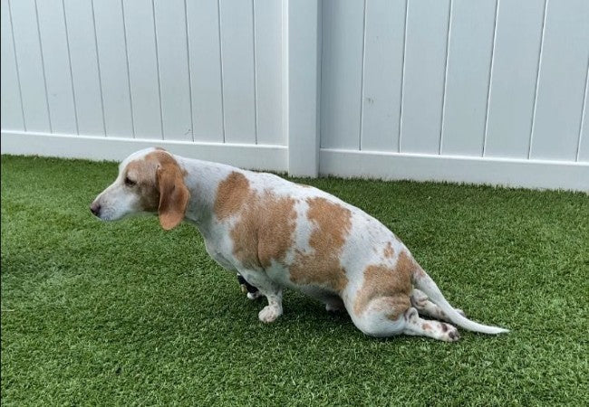
- Weakness or ataxia, especially in hind limbs; “knuckling” or dragging paws.
- Hunched posture, tense muscles, trembling.
- Severe cases: paralysis and loss of bladder/bowel control – urgent vet care needed.
🔍 Diagnostic Process
- Neurological & physical exam: Localizes injury, assesses pain, gait, and reflexes.
- X-rays: Reveal narrowed disc spaces and calcification—but don’t show the spinal cord.
- Advanced imaging (CT, myelography): Non-contrast CT/myelography has ~82–83% sensitivity; CT-myelography and MRI offer 94–97% accuracy in locating disc compression.
- MRI: Gold standard. T2-W images show spinal cord compression and hyperintensity; used to assess myelomalacia risk.
- CSF analysis: Rarely, it helps rule out inflammatory/spinal cord conditions.
🛠 Treatment Strategies
1. Conservative Medical Management
- Strict crate rest (4–6 weeks) to minimize movement.
- Anti‑inflammatories: NSAIDs (carprofen, meloxicam) or steroids for severe pain.
- Analgesics for comfort; muscle relaxants to ease spasms.
- Rehabilitation/physiotherapy: Strengthens core, restores function.
- Ideal for Type I dogs who can walk, and Type II mild to moderate disease.
2. Surgical Intervention
- Indicated for non-ambulatory dogs, loss of pain perception, or failing conservative treatment.
- Common procedures: Hemilaminectomy or dorsal laminectomy to remove compressive disc material.
- Higher success when performed before complete paralysis; mobility is often restored.
- Cost ranges: $2,000–$5,000 depending on location and complexity.
3. Rehabilitation & Long-Term Support
- Post-treatment rehab: hydrotherapy, core strengthening, gait retraining.
- Assistive devices: slings, carts for partial paralysis.
- Weight management and controlled activity help prevent recurrence.
- Preventative strategies: avoid jumping/dog stairs for predisposed breeds.
📈 Prognosis by Scenario
- Walking dogs with pain: Excellent (80–90%) recovery with conservative therapy.
- Surgical candidates: ~90% regain mobility if deep pain is intact; recovery is slower if surgery is delayed.
- Deep pain-negative: Guarded prognosis; early surgery and rehab improve chances.
- Type III/ANNPE: Many recover without surgery but benefit greatly from rehab.
🏡 Home Care & Prevention Tips
- Maintain a healthy weight—excess weight worsens spinal loading.
- Restricted environments—avoid stairs/jumping for at-risk breeds.
- Provide ramps for furniture and car access.
- Orthopedic dog beds and supportive harnesses reduce strain.
- Regular gentle exercise and controlled walking to strengthen the core.
📱 Ask A Vet Telehealth Support
- 📸 Upload videos/photos of gait, posture, and pain behaviors for remote evaluation.
- 🔔 Schedule and medication reminders during crate rest or post-op rehab.
- 🩺 Video consults to monitor recovery, adjust therapy, and troubleshoot complications.
🎓 Case Spotlight: “Rex” the Dachshund
Rex, a 6‑year‑old Dachshund, yelped after jumping into the car and developed pelvic limb weakness with intact pain perception. Diagnosis of T13–L1 Hansen Type I IVDD via MRI; hemilaminectomy performed within 24 hours. He began physiotherapy after 2 weeks and by 8 weeks was walking normally. Ask A Vet supported video check-ins, medication reminders, and delivered joint supplements. At 12 months post-op, Rex is active and pain-free. 🐕🦮
🔚 Key Takeaways
- IVDD is common in dogs, especially chondrodystrophic and large breeds.
- Signs range from pain to paralysis—rapid veterinary assessment is vital.
- Diagnosis uses X-ray, CT/myelography, and MRI MRI is most accurate.
- Treatment varies: crate rest and meds for mild cases; surgery in severe cases.
- Rehab and weight control improve outcomes and reduce future risk.
- Ask A Vet telehealth helps with remote monitoring, rehab guidance, medication delivery, and owner confidence 📲🐾
Dr Duncan Houston BVSc, founder of Ask A Vet. Download the Ask A Vet app today to support your dog through IVDD—from remote triage and diagnosis to post-treatment rehab, medication management, and home delivery 🐶📲


