

Veterinary Guide to Canine Iridociliary Cysts 2025 🩺🐶
In this article
Veterinary Guide to Canine Iridociliary Cysts 2025 🩺🐶
By Dr. Duncan Houston BVSc
🧬 What Are Iridociliary (Uveal/Iris) Cysts?
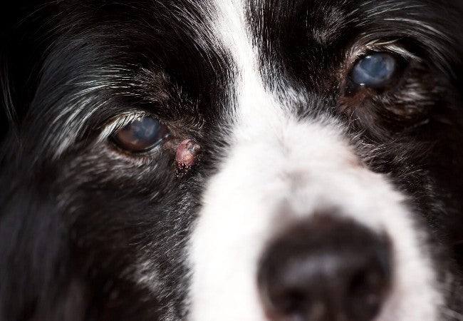
Iridociliary cysts are fluid-filled sacs that form within the iris or ciliary body, typically benign and often discovered incidentally during eye exams. They may float freely in the anterior chamber, adhere to the pupillary margin, or arise behind the iris. Appearance varies: pigmented (brown/black/yellow), smooth, circular, or oval.
👥 Who Is Affected?
- Age: Typically 7–9 years old, though it can occur at any age.
- Breeds: Golden Retrievers, Labrador Retrievers, Boston Terriers, Great Danes, and American Bulldogs are over-represented.
- Etiology: Can be congenital, spontaneous, traumatic, or inflammatory.
⚠️ Clinical Signs
- Often asymptomatic; discovered on routine eye exams or during mydriasis.
- Can cause “fly-biting” or vision obstruction if cysts float into the visual axis.
- Pigment deposits on cornea or lens capsule—common remnants of collapsed cysts.
- Rarely, large or multiple cysts may reduce aqueous outflow, elevating intraocular pressure (IOP), leading to glaucoma.
- Associated conditions in certain breeds: pigmentary uveitis in Goldens, BBBs; glaucoma in Bulldogs, Great Danes; cataract risk.
🔍 Diagnostic Approach
- Ophthalmic exam: Slit lamp/biomicroscopy reveals cyst size, shape, and location. Cysts transilluminate; tumors do not.
- Observe gravity effect: With head repositioning, free-floating cysts settle (“snow-globe” sign).
- Tonometry: Necessary if IOP is elevated or angle obstruction is suspected.
- Ultrasound biomicroscopy (UBM): Confirms cyst vs mass; defines ciliary body involvement.
- Ancillary imaging: High-frequency B-scan ultrasound, AS-OCT for detailed anatomy; MRI rarely needed.
🛠️ Management & Treatment
✳️ Observation (Preferred)
- Most cysts are benign—no treatment needed.
- Periodic exams (every 4–6 months) if cysts are large, pigmented, or in at-risk breeds like Goldens, Bulldogs, Great Danes.
⚠️ Intervention Indicated When:
- Cysts impair vision or cause “fly-biting” behavior.
- Obstruction of aqueous outflow or elevated IOP is detected.
- Signs of secondary uveitis or cataract formation.
🛠️ Treatment Options
- Laser therapy (diode/YAG): Least invasive; perforates cyst wall, drains fluid. Requires sedation or a topical anesthetic.
- Fine‑needle aspiration (FNA): Under magnification, drains cyst; small risk of inflammation, infection, cataract.
- Irrigation/aspiration: Automated surgical method for complete cyst removal via small incision.
- Intracystic sclerosis: Alcohol or antimitotic injection to collapse the cyst wall.
- Surgery: Reserved for deeply adhered or recurrent cysts not treatable via laser/FNA.
📈 Prognosis & Follow‑Up
- Excellent for benign cysts with no intervention.
- Cautions in predisposed breeds—monitor for uveitis, glaucoma, cataracts.
- Post-treatment rechecks: IOP, inflammation, recurrence at 1 week, 1 month, then every 6 months.
🏡 Home Care & Prevention
- Observe your dog’s vision—avoid bumping into objects, eye paw signs. Report changes.
- Administer prescribed anti‑inflammatory or pressure‑lowering drops post-procedure.
- Protect eyes from trauma—E-collar as needed.
- Adhere to the recommended schedule for eye exams, especially in breeds at higher risk.
📱 Ask A Vet Telehealth Support
- 📸 Upload crystal-clear “eye selfies” to assess cyst appearance, pupil reactions, and clarity remotely.
- 🔔 Get reminders for medication schedules, follow-up tonometry tests, and ophthalmologist referrals.
- 🩺 Video consultations to guide home eye drop technique and monitor recovery.
- 🧘 Educational content on laser vs aspiration treatments, monitoring techniques.
🎓 Case Spotlight: “Bailey” the Golden Retriever
Bailey, an 8-year-old Golden, was found to have multiple thin-walled iridociliary cysts and mild pigment deposits on the lens during a routine exam. Her IOP was normal, but she was referred to an ophthalmologist for baseline evaluation. No treatment was performed initially due to her lack of symptoms and free-floating nature. She was monitored every 6 months; after 18 months, one cyst drifted into her visual axis, causing a minor “fly‑biting” behavior. Laser drainage was performed, and topical NSAID drops were continued for two weeks. Follow-up showed resolution with no recurrence or inflammation. Ask A Vet helped schedule meds, provided drop reminders, and coordinated follow-up appointments. Bailey remains comfortable and vision-normal 🌟.
🔚 Key Takeaways
- Iridociliary cysts are benign fluid-filled sacs, common in middle-aged dogs, especially Goldens, Labs, Boston Terriers, and Great Danes.
- Typically asymptomatic and found incidentally; important to differentiate from uveal tumors using transillumination, slit-lamp, and UBM.
- Treatment is only if cysts impair vision, elevate IOP, or incite uveitis or cataracts.
- Management options: laser drainage, FNA, aspiration—choose the least invasive first.
- Prognosis excellent; breed-specific monitoring vital for early glaucoma or pigmentary uveitis.
- Ask A Vet telehealth supports remote assessment, medication guidance, specialist coordination, and peace of mind 📲🐾
Dr Duncan Houston BVSc, founder of Ask A Vet. Download the Ask A Vet app today to support your dog’s eye health—smooth remote triage, medication reminders, tele-guidance, and specialist referrals for the best vision care 🐶📲


