

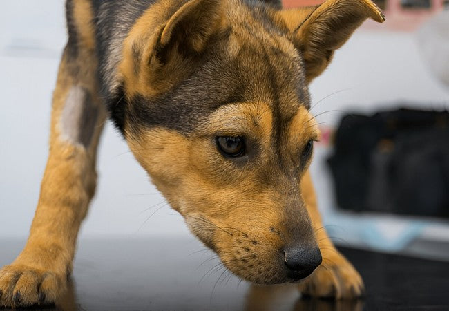
Veterinary Guide to Canine Systemic Vasculitis 2025 🩺🐶
In this article
Veterinary Guide to Canine Systemic Vasculitis 2025 🩺🐶
By Dr. Duncan Houston BVSc
🧬 What Is Systemic Vasculitis?
Systemic vasculitis is inflammation of blood vessel walls—arteries, veins, or capillaries—throughout the body. When immune complexes deposit or the endothelium becomes damaged, blood flow is compromised. This leads to visible skin signs and often affects organs such as the kidneys, liver, eyes, and nerves.
👥 Causes & Underlying Mechanisms
- Immune-complex hypersensitivity: Often type III reactions triggered by infection, drugs, food antigens, tick-borne diseases, neoplasia, or SLE.
- Drug or vaccine reaction: Onset 2–4 months after exposure—common with antibiotics or vaccines.
- Infection-induced: Bacterial, rickettsial (e.g., ehrlichiosis), viral, or fungal infections.
- Neoplastic triggers: Lymphoma, mast cell tumors via paraneoplastic vasculitis.
- Autoimmune diseases: SLE or systemic granulomatous vasculitis parallels human GPA.
👀 Clinical Signs & Systems Affected
🐾 Skin and Mucosa
- Petechiae, purpura, ecchymoses—especially on ears, tail, paw pads, lips.
- Vesicles, necrosis, and ulceration—leading to crusting or secondary infection.
- Swelling, firm edema (“pitting”), hair loss, and pain on palpation.
🌡️ Systemic & Organ Signs
- Fever, lethargy, weight loss, and anorexia indicate severe immune activation.
- Kidney involvement: proteinuria, hematuria, elevated BUN/creatinine.
- Neurologic deficits: seizures, meningitis, paresis if CNS vessels are affected.
- Ocular uveitis, hemorrhage; GI signs—vomiting, diarrhea from GI vessel inflammation.
🔍 Diagnostic Workup
- History: Recent meds, vaccines, potential infections, and tick exposure.
- Physical exam: Document skin lesions; palpate for pain, swelling; check lymph nodes, mucosal bleeding.
- Laboratory tests: CBC (leukocytosis, anemia), chemistry, urinalysis, tick titers, infectious panels.
- Skin and vessel biopsy: Gold standard—look for leukocytoclastic or granulomatous vasculitis.
- Imaging: Abdominal ultrasound, chest X-ray, MRI/CT if internal organs suspected.
- Special assays: ANA for SLE, ANCA in granulomatous forms (parallels human GPA).
🛠️ Treatment Plan
1. Remove Triggers
- Stop suspect drugs or vaccines—watch a 2–4 month resolution window.
- Treat infections: doxycycline or appropriate antibiotics/antifungals.
- Address neoplasia with an oncology consult if needed.
2. Immunosuppressive Therapy
- Glucocorticoids: Prednisone 2–4 mg/kg/day or methylprednisolone pulses for severe cases.
- Second-line: Azathioprine, cyclosporine, mycophenolate, or leflunomide in steroid-sparing roles.
- Severe cases: Cyclophosphamide (GPA‑like angiitis); consider plasma exchange in life-threatening organ involvement.
- Adjunctive agents: Pentoxifylline for microvascular circulation; topical tacrolimus for localized cutaneous lesions.
3. Supportive & Organ‑Specific Care
- Pain relief: NSAIDs initially, then taper before immunosuppression.
- Fluid therapy for renal support; electrolyte monitoring.
- Ocular treatment for uveitis—topical steroids and mydriatics.
- Neurologic monitoring and rehab support if CNS signs are present.
📈 Prognosis & Monitoring
- Prognosis depends on cause: idiopathic cutaneous forms are guarded to good with therapy; systemic/internal organ involvement is more serious.
- Concurrent vasculitis in pemphigus may delay remission and require prolonged therapy.
- Taper steroids when skin heals and labs normalize; reduce over months.
- Monitor CBC, chemistry, urinalysis monthly → every 3–4 months.
- Watch for relapse—a redeveloping rash, fever, or new systemic signs should prompt re-evaluation.
🏡 Home & Lifestyle Advice
- Prevent sun exposure—photosensitization can worsen lesions.
- Use soft bedding and minimize friction/trauma.
- Clean and medicate skin lesions as directed; monitor for infection.
- Maintain consistent medication schedules; never adjust without vet consultation.
- Track appetite, energy, lesions, urinary and neurologic changes daily.
📱 Ask A Vet Telehealth Support
- 📸 Upload rash and lesion photos; remote triage of flare vs. infection.
- 🔔 Medication tracker and taper reminders.
- 🩺 Lab schedule alerts—CBC, chemistry, urinalysis follow-ups.
🎓 Case Spotlight: “Hazel” the Rottie Mix
Hazel, a 5-year-old Rottweiler, had sudden purpura on her ears, footpads, and lethargy one week after a vaccine boost. Biopsy confirmed leukocytoclastic vasculitis. Treated with prednisone (3 mg/kg), doxycycline for tick prevention, and pentoxifylline. Skin healed in 10 days; prednisone tapered over 3 months. Two-year follow-up showed no relapse. Ask A Vet reminders kept her labs on track, and derm pics were monitored remotely. 🐕🌟
🔚 Key Takeaways
- Systemic vasculitis is immune-mediated vessel inflammation affecting skin & organs—early diagnosis is critical.
- Causes include drugs, infection, neoplasia, and autoimmunity; identification improves outcomes.
- Diagnosis relies on history, bloodwork, biopsy, and imaging.
- Treatment combines removal of triggers, corticosteroids, immunosuppressives, and organ support.
- Long-term monitoring and Ask A Vet telehealth support ensure safe tapering and relapse detection 🐾.
Dr Duncan Houston BVSc, founder of Ask A Vet. Download the Ask A Vet app today to receive remote guidance and support—photo triage, medication management, lab tracking, and home care to help your dog recover from vasculitis and stay well 🐶📲


