

Veterinary Guide to Eyeworm Infection in Dogs (Thelaziasis) (2025)🐶
In this article
Veterinary Guide to Eyeworm Infection in Dogs (Thelaziasis) (2025)🐶
By Dr. Duncan Houston BVSc
🔍 Introduction
Thelaziasis, commonly called “eyeworm infections,” is caused by parasitic nematodes (Thelazia spp.) infecting the conjunctival sac, tear glands, and ducts of dogs. These parasites, transmitted by tear-feeding flies, can cause eye irritation, ulcers, and—even rarely—blinding complications. Early detection and effective treatment are important for ocular health. 👁️
💡 What Are Eyeworms?
- Typically 10–15 mm long, pale white to cream nematodes visible on the eyeball, under eyelids, or within tear ducts.
- Common species infecting dogs include Thelazia callipaeda (Asia/Europe/US), T. californiensis (western North America), and T. gulosa (rare).
- Their life cycle involves tear-feeding flies that transmit infective larvae into the ocular tissues.
⚠️ Causes & Transmission
- Spread via non-biting flies (e.g. Phortica variegata, Fannia spp.) that feed on ocular secretions and deposit larvae.
- Dogs in endemic areas, with outdoor exposure, and those traveling are most at risk.
- Reported in Europe, parts of Asia, and emerging in the U.S. (e.g., New York).
🚨 Clinical Signs
- Excessive tearing (epiphora), conjunctivitis (redness, swelling), itchiness, and mucopurulent discharge.
- Frequent blinking, squinting, head shaking, and ocular discomfort.
- Severe cases may develop keratitis, corneal ulcers, opacity, or even vision impairment.
- Some dogs may show no symptoms despite harboring worms.
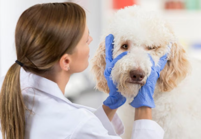
🔬 Diagnosis
- Primarily via ocular exam; worms may be visible on eye surface, under eyelids, or within tear ducts.
- Examination enhanced under topical anesthesia; sometimes saline flushing or magnification is needed.
- Ophthalmic evaluation rules out corneal damage and assesses tear production.
🛠 Treatment
- Mechanical removal: under local anesthesia, gently remove worms using forceps or flush with saline.
- Systemic/topical anthelmintics: ivermectin, moxidectin + imidacloprid, milbemycin oxime, or selamectin are effective.
- Anti-inflammatory/antibiotics: topical or systemic NSAIDs, corticosteroids, and antibiotics to manage conjunctivitis or keratitis.
📈 Prognosis & Follow‑Up
- Removal of worms plus medications leads to full recovery in most cases.
- Ulcers or corneal damage require extended monitoring and treatment.
- Recheck eyes 1–2 weeks post-treatment to verify worm clearance and healing.
🛡 Prevention
- Monthly prophylaxis with milbemycin oxime + afoxolaner (e.g., NexGard Spectra®) prevents reinfection in endemic areas.
- Other effective preventatives: spot-on ivermectin/moxidectin or oral milbemycin formulations.
- Minimize fly exposure—use screens and reduce breeding sites.
- Check eyes in dogs returning from travel to endemic regions.
🔧 Tools & Support Services
- Ask A Vet App: Immediate guidance on eye worm recognition, removal urgency, and treatment planning 📱
✅ Final Thoughts
Eyeworm infections may appear dramatic, but prompt mechanical removal, medication, and preventive care usually achieve full recovery. Prevention strategies—monthly parasiticides and reducing fly contact—are key in endemic zones. With tools like Ask AVet, dogs in 2025 can maintain clear, healthy eyes and comfortable vision. 🐾❤️
Download the Ask A Vet app today for expert help removing worms, choosing medications, and tracking eye wellness. 📱💡


