

Veterinary Guide to Hypopyon & Lipid Flare in Dogs 2025 🐶⚠️
In this article
Veterinary Guide to Hypopyon & Lipid Flare in Dogs 2025 🐶⚠️
By Dr. Duncan Houston BVSc
🔍 Introduction
Hypopyon is the accumulation of inflammatory cells (“pus”) in the eye’s front chamber, while lipid flare is a milky haze from blood-borne fats. These rare signs usually signal serious eye or systemic diseases. In this 2025 guide, I’ll explain their causes, recognition, diagnosis, treatments, and prevention. 🩺
💡 What Are Hypopyon & Lipid Flare?
- Hypopyon: Settling of white blood cells in the anterior chamber, forming a layer at the bottom.
- Lipid flare: Milky-white aqueous appearance due to hyperlipidemia combined with inflammation-based barrier breakdown.
⚠️ Causes & Risk Factors
- Severe anterior uveitis (infection, immune-mediated, trauma).
- Corneal ulcers or abscesses that trigger secondary inflammation.
- Neoplasia like ocular lymphoma—may contribute to hypopyon.
- Hyperlipidemia—raising blood lipid levels post-meal or chronically—enables lipid flare when uveitis is present.
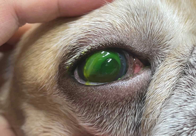
🚨 Clinical Signs
- Hypopyon: Yellow-white fluid accumulation in lower anterior chamber, often obscuring eye details.
- Lipid flare: Diffuse milky/cloudy appearance rather than a ventral layer.
- Both conditions may present with pain, squinting, tearing, red/blue cornea, blepharospasm, and vision loss.
- Signs of systemic illness may also be present if underlying disease like lymphoma or infection is involved.
🔬 Diagnosis
- Thorough ocular exam with slit lamp to visualize exudate and corneal changes.
- Differentiate by appearance: hypopyon forms a ventral layer; lipid flare is uniform and whiter.
- Tonometry to assess for secondary glaucoma risk.
- Work-up for underlying cause: blood work, urinalysis, infectious disease panels; imaging for masses.
- Anterior chamber paracentesis rarely recommended—can worsen inflammation.
🛠 Treatment
- Aggressive anti-inflammatory therapy: Topical/systemic corticosteroids or NSAIDs based on etiology.
- Antimicrobials: Based on suspected or confirmed infection—bacterial, fungal, viral, or parasitic.
- Lipid flare steps: Manage hyperlipidemia with diet, possibly systemic lipid-lowering strategies.
- Supportive measures: Atropine for pain/discomfort; monitor and treat intraocular pressure elevations.
- Surgery or biopsy: For suspected neoplasia—confirm via mass removal/histopathology.
📈 Prognosis & Follow-up
- Prognosis varies: guarded with neoplasia; better if the infectious/inflammatory cause is treated promptly.
- Secondary glaucoma and synechiae are possible if management delayed.
- Frequent rechecks: intraocular pressure, corneal health, inflammation status.
🛡 Prevention & Tips
- Keep up with regular eye exams and monitor any signs of uveitis or corneal disease.
- Manage lipid levels via diet or medication in hyperlipemic dogs.
- Treat eye trauma, infection, or inflammation promptly to prevent progression.
- Consider early specialist referral—ophthalmologist evaluation improves outcomes.
🔧 Recommended Tools & Services
- Ask A Vet App: 24/7 help for recognizing eye changes, medication use, and timing of referrals 📱
✅ Final Thoughts
Hypopyon and lipid flare are alarming ocular signs often indicating severe inflammation or systemic illness. Swift diagnosis, targeted treatment, and supportive care are essential to saving vision and overall health. Use reliable tools like AskAVet, manage eye conditions effectively in 2025 and beyond. 🐾❤️
Download the Ask A Vet app today for expert guidance on eye health, treatment plans, and monitoring—because every glance counts. 👁️📱


