

Veterinary Guide to Ulcerative Keratitis (Corneal Ulcers) in Dogs (2025)🐶
In this article
Veterinary Guide to Ulcerative Keratitis (Corneal Ulcers) in Dogs (2025)🐶
By Dr. Duncan Houston BVSc
🔍 Introduction
Ulcerative keratitis describes corneal ulcers—painful erosions that penetrate the epithelium and sometimes stroma. Prompt diagnosis and tailored treatment protect your dog’s vision and comfort in 2025 and beyond. 👁️
💡 What Are Corneal Ulcers?
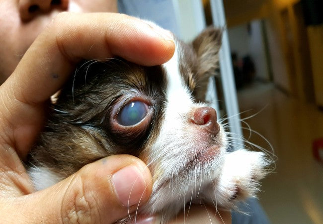
- Superficial erosions affect epithelium only; deeper stromal ulcers penetrate into the stroma; descemetoceles reach Descemet’s membrane.
- Fluid accumulates within the stroma, causing corneal opacity and pain.
⚠️ Common Causes
- Trauma: scratches from grass, nails, sticks, chemical exposure.
- Pre-existing issues: KCS (dry eye), dystrophy, entropion, distichiasis, endocrine diseases.
- Infections: bacterial, fungal, or immune-mediated conditions.
🚨 Clinical Signs
- Severe ocular pain: squinting, blinking, pawing at eye, photophobia.
- Discharge, corneal haze, redness, blood vessel growth.
- Miosis due to reflex uveitis; deeper ulcers may perforate and lead to vision loss.
🔬 Diagnostic Steps
- Fluorescein stain: highlights ulcers by dye retention.
- Evaluate tear production (Schirmer test), corneal sensitivity, eyelid conformation, and rule out underlying eyelid or systemic issues.
- Culture or cytology in complicated or infected ulcers.
- Monitor healing progress with repeat staining every 1–7 days.
🛠 Medical Management
- Topical antibiotics: broad-spectrum drops/ointments—every 4–6 hrs for superficial; every 2–4 hrs for complicated ulcers.
- Pain control: topical atropine for ciliary spasm; NSAIDs systemic as needed.
- E-collar: essential to prevent self-trauma.
- Debridement: grid or punctate keratotomy for non-healing ulcers.
🔧 Surgical Interventions
- Conjunctival flap/autograft: covers deep/stromal ulcers or descemetoceles to aid healing.
- Corneal debridement: removes necrotic tissue supporting healing.
- Bandage contact lens: protects the ulcer and maintains the tear film.
📈 Prognosis & Follow‑Up
- Superficial ulcers typically heal in 5–7 days; deep or melting ulcers require ongoing monitoring and follow-up.
- Potential complications: scarring, neovascularization, uveitis, glaucoma, vision loss.
- Regular rechecks during treatment; adapt therapy based on healing pace.
🛡 Prevention & Owner Advice
- Prevent ocular trauma—avoid unsafe play and clear the eye area of sharp objects.
- Maintain eyelid health—treat entropion, distichiasis, and dry eyes early.
- Use artificial tears routinely in dogs predisposed to KCS or ocular dryness.
- Monitor high-risk dogs (e.g., Boxers, brachycephalics) closely after eye irritation.
🔧 Tools & Support Services
- Ask A Vet App: 24/7 support for ulcer detection, medication planning, and emergency referrals 📱
✅ Final Thoughts
Corneal ulcers are painful and vision-threatening. With timely diagnosis, aggressive medical/surgical care, and preventive strategies, many dogs fully recover. Using Ask AVet, vigilant follow-up and ocular health into 2025 and beyond. 🐾❤️
Download the Ask A Vet app today for expert ocular care support—ulcer monitoring, medication guidance, and vision protection. 📱💡


