


Vet’s 2025 Guide to Granulomatous Meningoencephalitis (GME) in Dogs – Causes, Diagnosis & Treatment 🧠🐾
By Dr. Duncan Houston BVSc
💡 What Is GME?
GME is an immune-mediated, idiopathic inflammatory disease of the central nervous system, typically affecting the brain, spinal cord, and meninges in dogs. It may present as disseminated (widespread inflammation), focal (localized lesions resembling tumors), or ocular (optic nerve involvement) forms.
⚠️ Who’s at Risk?
- Small breed and toy dogs—especially Poodles, Terriers, Chihuahuas, Yorkshire, Maltese, Dachshunds
- Typically young to middle-aged, with female dogs slightly more affected
🧩 Key Symptoms
- Seizures, altered behavior or mentation, head tilt
- Ataxia, loss of coordination, weakness, neck pain, paralysis
- Vision loss in ocular form, circling or blindness in focal disease
🔍 Diagnosis
- MRI: reveals focal or multifocal hyperintense T2/FLAIR brain lesions with mass effect and contrast enhancement
- CSF analysis: shows mononuclear pleocytosis—high lymphocytes/monocytes/protein
- Biopsy: confirms diagnosis via perivascular granulomatous inflammation, but often not pursued in live dogs
💊 Treatment Options
GME requires aggressive immunosuppression and long-term management:
- High-dose corticosteroids (e.g., prednisone) initially
- Additional immunosuppressives—azathioprine, cyclosporine, cytarabine, methotrexate—often used together
- Focal GME may respond well to radiation therapy alongside drugs
🛟 Supportive & Long-Term Care
- Seizure control—anti-epileptics (e.g., phenobarbital, zonisamide)
- Pain management and physical support (e.g., nonslip mats, ramp access)
- Ask A Vet helps with monitoring complex drug protocols and symptom changes
- Controlled environment to ensure safety during ataxia or seizures
📈 Prognosis & Outlook
- Acute cases may progress rapidly—without treatment, many dogs decline in days to weeks
- With aggressive therapy, some dogs achieve remission for months to years—median survival varies widely
- Prompt treatment and consistent follow-up improve outcomes
🛡️ Prevention & Monitoring Tips
- Early neurologic evaluation for small-breed dogs with sudden neuro signs
- Sequential MRI/CSF tests monitor treatment response and relapse
- Owner vigilance—immediate veterinary contact for new seizures or ataxia
📲 Owner Support Tools
- Ask A Vet: Immediate advice on medication dosing, seizure monitoring, side effects
🌟 Case Snapshot
Case: A 5-year-old Miniature Poodle showed seizures and circling. MRI revealed a focal temporal lobe lesion; CSF confirmed GME. Treated with prednisone + cyclosporine + radiation. The dog remains seizure-free at 14 months with tapered meds and regular MRI/CSF monitoring. 🧠👏
✅ Final Takeaways
- GME is a serious, immune-mediated neurologic disease common in small breeds
- Diagnosis relies on MRI and CSF; biopsy confirms the disease
- Requires aggressive immunosuppression and possibly radiation therapy
- Supportive care and monitoring are critical for long-term management
- Ask A Vet provides vital help with neurologic and medication coordination 🧠
📥 Immediate Support
If your dog shows seizures, ataxia, head tilt or neurologic changes, download the Ask A Vet app for urgent consultation and care guidance. Visit AskAVet.com for 24/7 expert support. 🐾🩺
This article is general veterinary education and does not diagnose or treat an individual animal. Always confirm medication names, doses and interactions with the veterinarian who knows your pet.
If your animal is struggling to breathe, collapsed, bleeding heavily, seizing, or has eaten something toxic, contact your vet or the nearest emergency clinic now.
Animal Poisons Helpline (AU) 1300 869 738 · Animal Poisons Helpline (NZ) 0800 869 738 · ASPCA Animal Poison Control (US) (888) 426-4435
More from Ask A Vet
For everyday pet care, one scan or tap of the smart pet ID tag opens your pet's free profile on any phone, no app needed; the GPS pet tracker logs live location and activity trends; and the Ask A Vet app keeps your pet's records and routines together.
 QR Code Dog & Cat ID Tag with NFC, HangingView ID Tag
The ASK A VET™ Smart ID Tag is a QR scan or NFC tap on any phone, no app needed, with a free profile you can update anytime.
 Dog & Cat GPS Tracker for Their CollarView tracker
The ASK A VET™ Tracker logs live location and activity trends, so a change in routine never goes unnoticed.
 Talk to a Vet Online: The Ask A Vet AppExplore the app
Ask questions, speak with vets, and keep your pet's records and routines together with Ask A Vet.
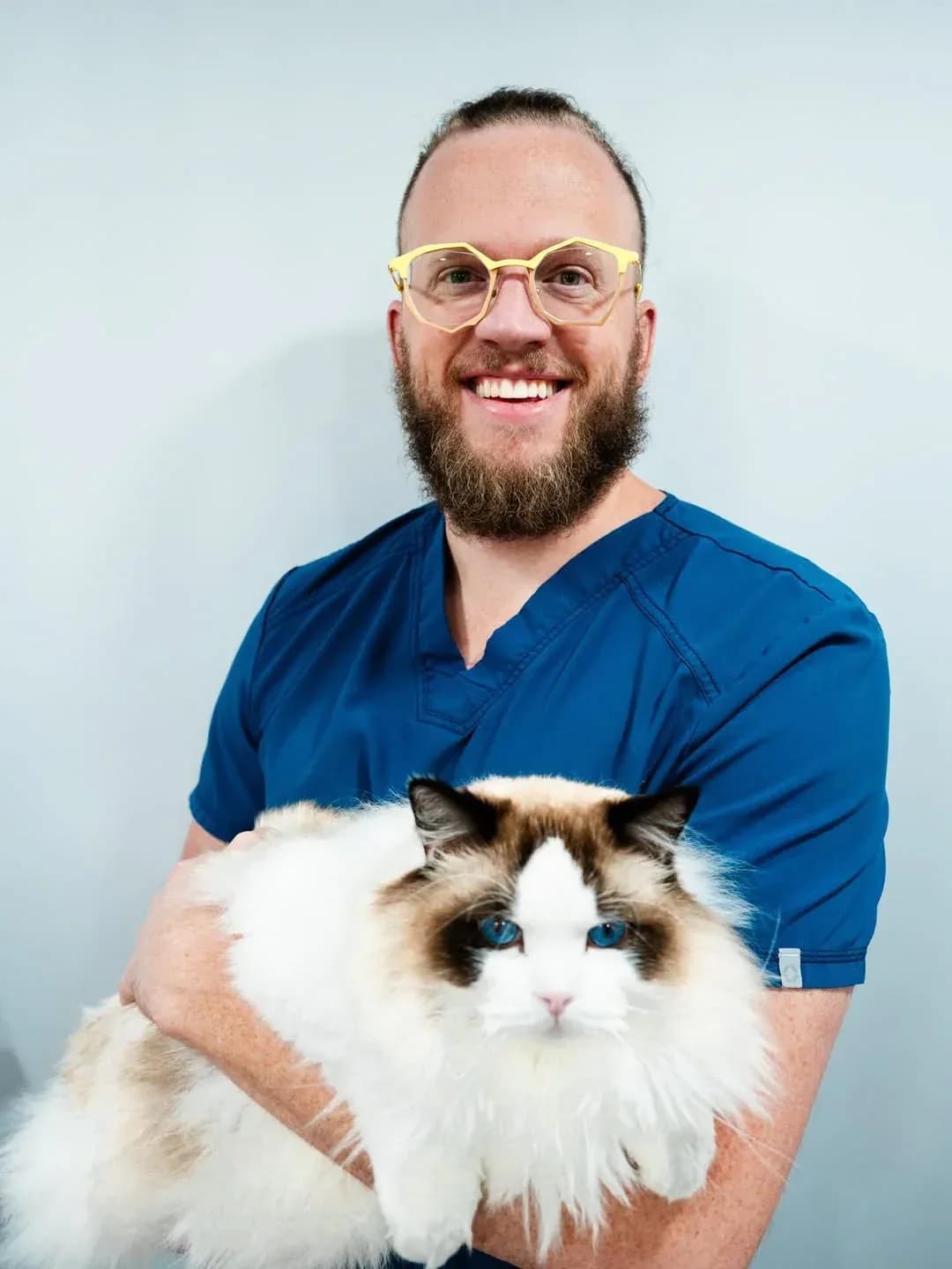
Ask A Vet articles are written by Dr Duncan Houston BVSc, a practising veterinarian, unless another author is named in the byline.